
Fine motor skill training enhances functional plasticity of the corticospinal tract aTher spinal cord injury
2017-01-21 03:33:12JianLiuXiaoyuYangWeiweiXiaJianDongMaoguangYangJianhangJiao
中國神經(jīng)再生研究(英文版) 2016年12期
Jian Liu, Xiao-yu Yang, Wei-wei Xia, Jian Dong, Mao-guang Yang, Jian-hang Jiao
Department of Orthopedics, China-Japan Union Hospital, Jilin University, Changchun, Jilin Province, China
Fine motor skill training enhances functional plasticity of the corticospinal tract aTher spinal cord injury
Jian Liu, Xiao-yu Yang*, Wei-wei Xia, Jian Dong, Mao-guang Yang, Jian-hang Jiao
Department of Orthopedics, China-Japan Union Hospital, Jilin University, Changchun, Jilin Province, China
How to cite this article:Liu J, Yang XY, Xia WW, Dong J, Yang MG, Jiao JH (2016) Fine motor skill training enhances functional plasticity of the corticospinal tract aTher spinal cord injury. Neural Regen Res 11(12):1990-1996.
Open access statement:This is an open access article distributed under the terms of the Creative Commons Attribution-NonCommercial-ShareAlike 3.0 License, which allows others to remix, tweak, and build upon the work non-commercially, as long as the author is credited and the new creations are licensed under the identical terms.
Funding:This study was supported by the National Natural Science Foundation of China, No. 30972153.
Graphical Abstract
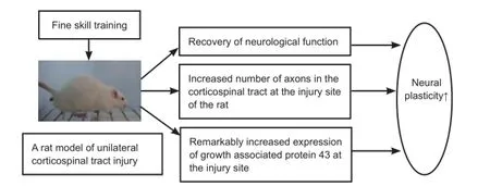
Fine motor skill training promotes functional recovery and enhances neural plasticity following unilateral corticospinal tract injury
Following central nervous system injury, axonal sprouts form distal to the injury site and extend into the denervated area, reconstructing neural circuits through neural plasticity. How to facilitate this plasticity has become the key to the success of central nervous system repair. It remains controversial whether fne motor skill training contributes to the recovery of neurological function aTher spinal cord injury. Therefore, we established a rat model of unilateral corticospinal tract injury using a pyramidal tract cutting method. Horizontal ladder crawling and food ball grasping training procedures were conducted 2 weeks before injury and 3 days aTher injury. The neurological function of rat forelimbs was assessed at 1, 2, 3, 4, and 6 weeks aTher injury. Axon growth was observed with biotinylated dextran amine anterograde tracing in the healthy corticospinal tract of the denervated area at diferent time periods. Our results demonstrate that compared with untrained rats, functional recovery was better in the forelimbs and forepaws of trained rats. The number of axons and the expression of growth associated protein 43 were increased at the injury site 3 weeks aTher corticospinal tract injury. These fndings confrm that fne motor skill training promotes central nervous system plasticity in spinal cord injury rats.
nerve regeneration; spinal cord injury; plasticity; axons; functional training; corticospinal tract; growth associated protein 43; neural regeneration
Introduction
The central nervous system can regulate itself to adapt to environmental changes or injuries through neural plasticity (Celnika and Cohen, 2004). Far from the injured area neural plasticity forms axon collaterals, whose growth and extension are not afected by the inhibitory environment at the injury site. However, axon regeneration and extension in adult human and animal central nervous systems is limited.Thus, the facilitation of neural plasticity has become the key to treating central nervous system injury.
Neural plasticity can be enhanced through functional training to promote functional recovery aTher central nervous system injury. Previous studies have shown that cerebral cortex size, the number of dendritic branches, and neurogenesis in the hippocampus are improved in rodents kept in environments that enhance motor activity or improve sensory and cognitive abilities (Girgis et al., 2007; Maier et al., 2008).These enhanced environments can promote the recovery of motor function aTher spinal cord injury and stroke (Z’Graggen et al., 1998; Shibolet et al., 2004). Sensory stimulationand targeted exercises effectively restore motor function after brain injury (Whishaw et al., 1993). Nevertheless, these interventions are more biased towards particular body movements or autonomous mechanical movements.The efect of fne motor skills on functional recovery aTher spinal cord injury is still controversial. Metz and Whishaw (2000) stated that functional recovery after central nervous system injury needs special post-traumatic functional training, such as grasping movements. Even slight recovery in hand function can noticeably improve patient’s quality of life aTher spinal cord injury (Metz and Whishaw, 2000; Bolton et al., 2006).
Therefore, functional training has an important infuence on the regulation of axonal growth, through the extension and guidance of axons aTher spinal cord injury. We investigated whether fne motor skill training can enhance neural plasticity and promote the recovery of neurological function aTher spinal cord injury in rats.
Materials and Methods
Experimental animals
One hundred and twenty-eight female Sprague-Dawley rats weighing 220-250 g were obtained from the Experimental Animal Center of Jilin University of China [license No. SCXK (Ji) 2008-0005]. Of them, 56 underwent neurological assessment and axon counting, and 72 were used for western blot assays. The rats were housed in individual cages under a 12-hour light/dark cycle in a dry and ventilated room at 23-25°C, with free access to food and water. All surgery was performed under anesthesia, and all efforts were made to minimize pain and distress in the animals. All procedures were carried out in accordance with the United States National Institutes of Health Guide for the Care and Use of Laboratory Animal (NIH Publication No. 85-23, revised 1986). The study was approved by the Animal Ethics Committee of Jilin University of China.
Preparation of rat models of unilateral corticospinal tract injury
Fifty-six rats were randomly assigned to seven groups (eight per group). The unilateral corticospinal tract injury model was established as follows: The rats were intraperitoneally anesthetized with 10% chloral hydrate, and fxed on the table in the supine position. The skin of anterior portion of the neck was incised, and muscles were dissociated. The trachea and esophagus were moved to the right to expose the occipital bone. The occipital bone at the basilar part was removed by grinding with a mill, and the vertebral body was exposed. A custom-made crochet was inserted into the vertebral body with a depth of 0.1 cm and the leTh pyramidal tract was resected. ATher thorough hemostasis, the incision was sutured. The rats were placed on an electric blanket until regaining consciousness.
In the sham group, the occipital bone at the basilar part was removed and the centrum exposed, but the pyramidal tract was not injured.
In the untrained group, the unilateral corticospinal tract was injured for 1 week, without any training. In the 1-, 2-, 3-, 4-, and 6-week unilateral corticospinal tract injury groups, fne motor skill training was conducted before and aTher unilateral corticospinal tract injury.
Fine motor skill functional training and functional behavioral assessment
Horizontal ladder crawling and food ball grasping were performed and functioning was evaluated prior to sample collection at each time point. Each rat was scored three times in each test, and the average value was recorded.
(1) Food ball grasping
A transparent resin training box (45 cm high, 12.5 cm wide and 38.5 cm long) was as described in a previous study (Whishaw et al., 1993). The bottom of the box was constructed of hollow mesh and raised 3 cm from the ground to ensure the rats could not pick up falling food. A narrow gap (1 cm wide and 10 cm high) on the side wall of the box enabled the rats to grasp the food balls with their forelimbs. The food balls outside the side wall were placed 3 cm from the bottom of the box. The distance between the ball and the inner wall of the box was 2 cm to prevent the rat from passing the ball into the interior of the box with its tongue. If the rats could retrieve and eat the food balls, the experiment was recorded as successful. If the rats were unable to retrieve the food balls or the food balls through the bottom of the cage, the experiment was recorded as failure.
Training methods: The rats received food ball grasping training, 6 days per week, 2 weeks before injury, until reaching a success rate of at least 65%. The food ball grasping assessment commenced 3 days after injury, for 6 days per week. According to the group assignment, functions were scored the day before sample collection. In the untrained group, functioning was scored 6 weeks aTher injury and before sample collection.
Scoring criteria: In accordance with a previous study (Metz and Whishaw, 2000), the process of grasping food balls was divided into seven actions. (1) Extending the forelimbs: Forelimb and elbow were extended towards food balls. (2) Separating forepaws: The forepaw was separated and placed over the surface of the ball. (3) Forepaw palm down: Elbow stretched out. The forepaw was placed on the surface of the ball and scratched it. (4) Grasping: The rat forepaw completely grasped the ball. (5) Intorsion I: Elbow intorsion and forepaw retraction, during which the forepaw intorsion was 90°. (6) Intorsion II: The forepaw retracted and moved to the rat’s mouth. (7) Releasing action: The rat sat and used both forepaws to put food into their mouth. Each action was recorded in accordance with three grades: If the action occurred, the score 1 recorded; if not, 0. If the action occurred, but the performance was abnormal, the score was recorded as 0.5. If an action did not occur, this did not necessarily mean that the grasping process stopped, as there may be other compensatory actions allowing completion of the process. The average value of three grasps was recorded for each rat.
(2) Horizontal ladder crawling
A horizontal ladder (1 m long) was made in accordance with a previous study (Bolton et al., 2006). An iron bar was placed along horizontal direction. To avoid the rats becoming familiar with the iron rod spacing, the iron rods were placed freely in the range of 1.5-3.0 cm. The horizontal ladder was 30 cm from the ground.
Training methods: The rats received horizontal ladder crawling training at 2 weeks before injury, three times every other day. The training assessment commenced 3 days aTher injury, three times every other day. According to the group assignment, the success rate was recorded the day before sample collection (Figure 1). The success rate was equal to normal gait/all gait × 100%. In the untrained group, the success rate was recorded 6 weeks after injury and the day before sample collection.
Biotinylated dextran amine (BDA) anterograde tracing
All rats were injected with BDA 1 week before injury. In accordance with a previous study (Hendriks et al., 2006), the rats were fixed on a stereotaxic apparatus after anesthesia as mentioned above (Figure 2). After shaving and sterilizing, the midline scalp was incised, and the skull was exposed. The center of the anterior fontanel was considered the marker. A sagittal midline incision was made along the sagittal suture. Eight holes approximately 0.5 mm diameter were drilled with a mill on each side (coordinates: 1.0 mm anterior to the center of anterior fontanel, 1.0 mm posterior to the center of anterior fontanel and midline of anterior fontanel, 2.0 mm posterior to the center of anterior fontanel, and 1.5 mm left and 2.5 mm right to the midline). A microsyringe was fixed on the stereotactic injection support, with the injection points above the holes. BDA-10000 10% (Vector Laboratories, San Francisco, CA, USA) was slowly injected in each hole, 0.2 μL at a time. BDA was injected separately at 1.0 and 2.0 mm depths from the cerebral cortex at each injection point. That is, the needle was maintained in place for 1 minute aTher injection at 2.0 mm depth, and then withdrawn slowly. The needle was then maintained in place for 5 minutes aTher injection at 1.0 mm depth. There were 32 injection points in each rat. The total dose of BDA was 6.4 μL.
Morphology of axons
Tissue preparation: ATher training, the rats were anesthetized using the method described above. The heart was perfused with 4°C physiological saline and 0.1 M phosphate bufered saline (PBS) (pH 7.4) containing 4% paraformaldehyde. The spinal cords (C6-T1) from the brain stem to the enlarged cervical segment were fxed in 0.1 M PBS (pH 7.4) containing 4% paraformaldehyde at 4°C overnight, and placed in 0.1 M PBS containing 30% sucrose for 5 days until the tissues sank.
Visualization with 3,3′-diaminobenzidine (DAB): The spinal cords (C6-T1) were embedded with frozen embedding media, and axially sliced into 40 μm-thick frozen sections with a freezing microtome. Sections were washed for 3 minutes three times with Tris-bufered saline with Tween (TBST) supplemented with 50 mM Tris, 0.9% NaCl, 0.5% Triton X-100, pH 8.0. These sections were then incubated with avidin-biotin-peroxidase complex (Vectastain ABC Elite kit; Vector Laboratories; 1:100 in TBST) at 4°C overnight, then washed for 3 minutes three times with TBST, followed by a wash with 50 mM Tris-HCl (pH 8.0). Subsequently, sections were incubated with 0.4% ammonium nickel sulfate (Sigma, St Louis, MO, USA), and with a mixture of ammonium nickel sulfate and 0.015% DAB (Sigma). The sections were treated with a mixture of 0.4% ammonium nickel sulfate, 0.015% DAB and 0.004% H2O250 mM Tris (pH 8.0) for 5-10 minutes. The reaction was terminated with 50 mM Tris-HCl. When dry the sections were mounted.
Quantitation of corticospinal tract axons in the midline of the spinal cord
Axonal sprouting and elongation in 40 frozen serial spinal cords sections (C6-T1) were observed with an optical microscope (Olympus, Tokyo, Japan). This segment of spinal cord was chosen because the motor neurons in C6-T1dominate the fine motor movement of the forelimbs (Maegele et al., 2005). The axons elongated from collateral sprouting presented with very irregular extension in the gray matter. To avoid repeat counting, axons traversing I, II, or III on each slice were used for quantitation (Figure 3). Serial sections of the pyramidal tract of the brain stem of each rat were made to allow for the correction of anomalies caused by individual diferences in sensorimotor cortex BDA uptake. Axons of three rectangular areas on each slice of the four serial sections were quantifed, and the average value was calculated. The relative number of corticospinal tract axons traversing the midline of the spinal cord was the number of axons traversing I, II, and III of each rat/the average number of axons in the corresponding vertebral body (Figure 3).
Western blot assay of GAP-43 expression in the rat spinal cord
The remaining 72 rats were randomly assigned to three groups. The control group rats (n = 24) received no intervention. In the trained group (n = 24), rats underwent unilateral corticospinal tract injury and functional training. In the untrained group (n = 24), rats only received unilateral corticospinal tract injury and no training. Each group was subdivided into 1-, 2-, 3-, and 4-week post-injury groups (n = 6). The model establishment and training methods were identical to those described above. At the appropriate time points, rats were intraperitoneally anesthetized with 10% chloral hydrate (30 mg/kg). An incision was made through the posterior approach to the cervical spine. ATher removal of the C1-T2vertebral plate, the spinal cord was exposed and the bilateral nerve root resected. C4-T1spinal cord on the healthy side was obtained, washed with frozen physiological saline, and stored in liquid nitrogen for western blot assay. After sodium dodecyl sulfate polyacrylamide gel electrophoresis, proteins were transferred onto membranes. The membranes were blocked with phosphate buffered salinewith Tween (PBST) containing 5% defatted milk powder for 90 minutes, washed three times with PBST, incubated with growth associated protein 43 (GAP-43) and β-actin antibodies at room temperature for 30 minutes, and then placed on a shaking table at 4°C overnight. The membranes were then incubated with IgG antibody (1:1,000) on a shaking table at room temperature for 90 minutes, washed three times with PBST, visualized with 3,3′-diaminobenzidine, and exposed to X-ray flm. Results were expressed as the optical density ratio of the target protein to β-actin.
Statistical analysis
Measurement data, expressed as means ± SD, were analyzed using SPSS 13.0 software (SPSS, Chicago, IL, USA). The differences between groups were compared using one-way analysis of variance and Fisher’s least significant difference post hoc test. A value of P < 0.05 was considered statistically signifcant.
Results
Identifcation of rat models of unilateral corticospinal tract injury
BDA anterograde tracing results revealed that model achieved the requirement of unilateral corticospinal tract injury (Figure 4).
Efects of fne motor skill training on motor function
Horizontal ladder crawling and food ball grasping results demonstrated that functional scores were signifcantly diferent in each corticospinal tract injury group compared with the sham group (P < 0.01). In the 1-, 2-, 3-, 4-, and 6-week unilateral corticospinal tract injury groups, motor function gradually recovered within 3 weeks of the injury, then plateaued. Motor functions in the untrained and 1- and 2-week unilateral corticospinal tract injury groups were signifcantly diferent compared with the 3-week unilateral corticospinal tract injury group (P < 0.01). Motor function was not signifcantly diferent in the 3-, 4-, and 6-week unilateral corticospinal tract injury groups (Figure 5).
Efects of fne motor skill training on spinal cord axon numbers
ATher absorption by neuronal cells in the rat cerebral cortex, BDA was used as an anterograde tracer along the corticospinal projection pathway. Approximately 95% of corticospinal tracts enter the deep part of posterior funiculus of the contralateral spinal cord through pyramid decussation to dominate fine movement of the lateral limb, especially the forelimb. Our results revealed that BDA was found within the rat cerebral cortex, and was well absorbed. This elucidates the corticospinal projection pathway and the sprouting and elongation of axon collaterals. Due to the small size of the spinal cord, thick sections, and irregular axon growth, collateral sprouting was difcult to observe clearly under the microscope. Thus, images of each part of each section were seamlessly stitched with Photoshop CS5 software (Adobe Systems, San Jose, CA, USA) to display the complete processof axonal growth. This was drawn on the picture of the spinal cord that was manually drawn with a pencil to illustrate the growth of the axons on the injured side during diferent time periods. We found that the number of axons gradually increased and extended long distances within 3 weeks of the injury. Three weeks postoperatively, the number and length of axons tended to be stable. Motor functions in the sham, untrained, and 1- and 2-week unilateral corticospinal tract injury groups were signifcantly diferent as compared with the 3-week unilateral corticospinal tract injury group (P <0.01). There were no significant differences in motor function between the 3-, 4-, and 6-week unilateral corticospinal tract injury groups (Figure 6).
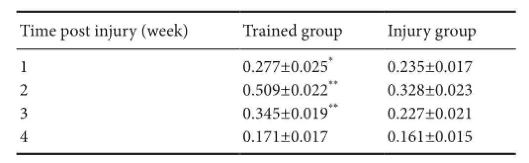
Table 1 Growth associated protein-43 expression in the spinal cord of rats with corticospinal tract injury at various time points
Efects of fne motor skill training on GAP-43 expression
Western blot assay results revealed that GAP-43 was expressed in the rat cervical cord of the control group, but its expression was low. GAP-43 expression could be detected in injured cervical cords at 1 week. There were signifcant differences in GAP-43 expression between the injury group and the normal group (P < 0.05). GAP-43 expression reached a peak at 2 weeks, gradually reduced at 3 weeks, and was close to control level at 1 week. No signifcant diference in GAP-43 expression was seen between the injury and normal control groups at 4 weeks (P > 0.05). In the trained group, GAP-43 expression appeared at 1 week, peaked at 2 weeks, and gradually decreased at 4 weeks to close to that of the control group (P > 0.05). GAP-43 expression was significantly higher at 1, 2, and 3 weeks aTher injury in the trained group compared with the injury group (P < 0.05). Moreover, significant differences in GAP-43 expression were identified between the trained group at 2 and 3 weeks and the injury group (P < 0.01; Figure 7, Table 1; data not shown for the normal control group).
Discussion
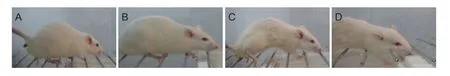
Figure 1 Behavioral changes in rats during horizontal ladder crawling.
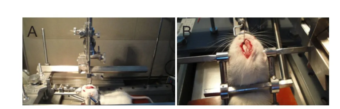
Figure 2 A rat fxed on the stereotaxic apparatus for biotinylated dextran amine anterograde tracing.
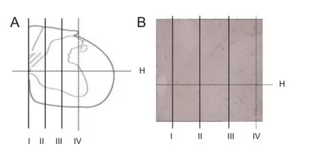
Figure 3 Quantitation of axons in the corticospinal tract traversingthe midline of the spinal cord.

Figure 4 Biotinylated dextran amine anterograde tracing of rat unilateral corticospinal tract (3,3′-diaminobenzidine staining, light microscope, ×100).
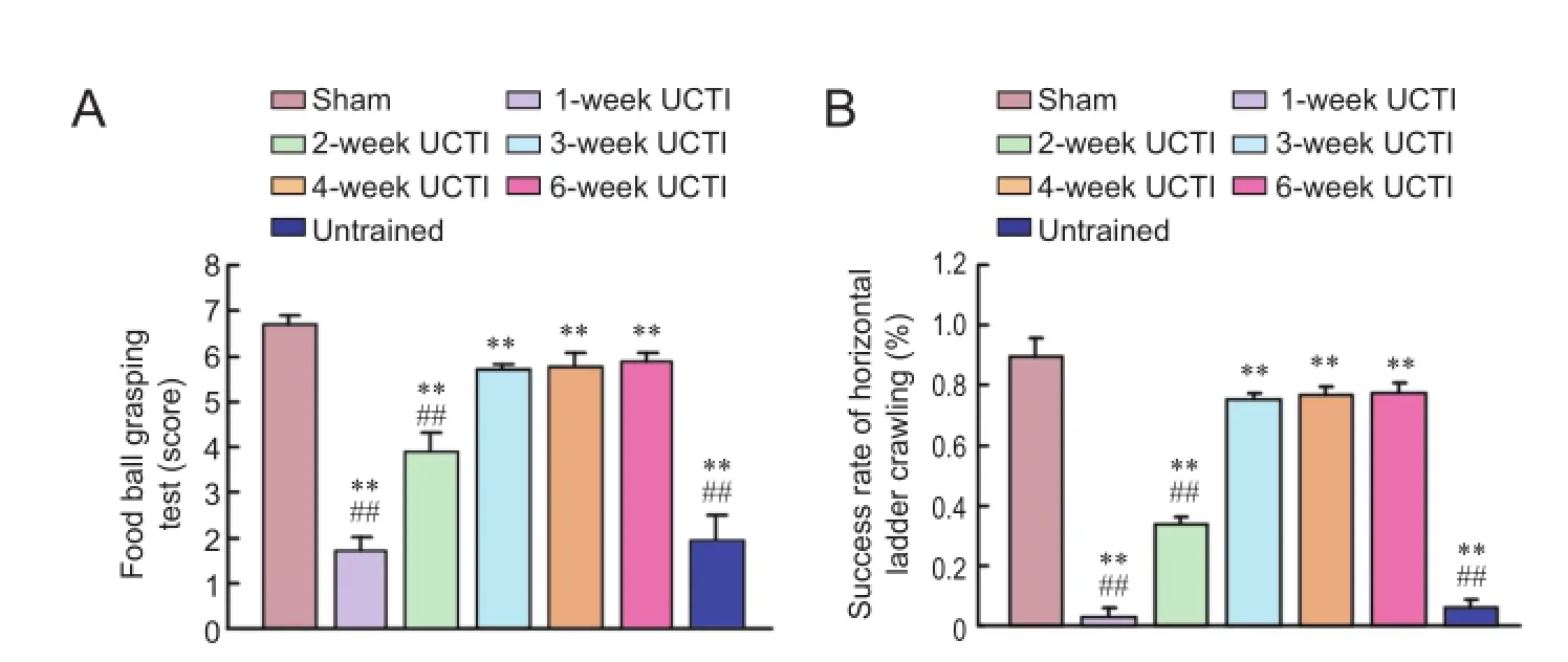
Figure 5 Efects of fne motor skill training on motor function in a rat model of unilateral corticospinal tract injury.
The corticospinal tract originates in the sensorimotor cortex and is the main motor tract within the spinal cord of mammals (Jang et al., 2015). Its fibers innervate spinal anterior horn cells directly, or indirectly through intermediate neurons, to control the movement of skeletal muscle. The rat corticospinal tract resides in the deepest layer of the dorsal funiculus of the spinal cord. The fiber bundle is small and deep and the currently used method of spinal cord hemitransection makes it difcult to ensure complete transection of the corticospinal tract (Hendriks et al., 2006). Moreover, the establishment of spinal cord hemitransection models needs to remove the lamina and to expose the spinal cord.The technique has many disadvantages, including a complicated local structure, difculty of operation, large degree of traumatic injury, difcult care aTher injury, low success rate, and other infuencing factors. Studies of the mechanism of spinal cord regeneration are limited. Through anatomical observation of the rat corticospinal pathway, according to morphological characteristics and anatomical localization of corticospinal tract, one side of the pyramidal tract is selectively dissociated. A model of small-range selective corticospinal tract injury is needed to provide a simple, reliable, and practical animal model for studying the regeneration and reconstruction of the central nervous system.
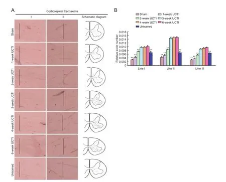
Figure 6 Efects of fne motor skill training on the number of axons in a rat model of UCTI.
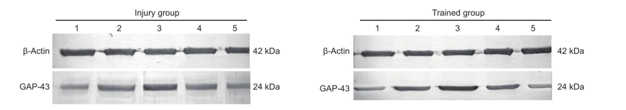
Figure 7 Efects of fne motor skill training on GAP-43 expression in a rat model of unilateral corticospinal tract injury.
Results have confrmed that functional recovery requires special post-traumatic functional training aTher central nervous system injury. In particular, the recovery of fetching functioning aTher spinal cord injury requires grasping training. We found that the three weeks after injury is the key time window for facilitating changes in neurological functional plasticity. This time window provides the experimental basis for subsequent studies of the sprouting and elongation of corticospinal tract axons and the precise mechanisms of neural plasticity.
BDA anterograde tracing demonstrated that with three weeks of injury was the key time period for axonal growth and synapse formation. One week aTher injury, some axons passed through line II, but there were fewer formed synapses. Therefore, the newly formed axons did not bind well to the dendrites of neuronal cells in spinal gray matter to form synapses. The neural circuits were not fully formed and so the functional recovery was not remarkable. Two to three weeks after injury, axons not only passed through the line II, but many synapses were also formed with an increase in functioning also. Three weeks later, the numbers of axonsand synapses stopped increasing and functional recovery plateaued. At this time point, forelimb and forepaw functioning was still noticeably different to that of rats in the sham group.
Our results demonstrate that changes in GAP-43 expression are consistent with BDA anterograde tracing. This suggests that functional training can efectively promote axonal sprouting and extension aTher corticospinal tract injury, and the formation of new neural circuits. Therefore, it may be one of the efective ways to promote neural plasticity.
The injection of BDA in the cerebral cortex caused substantial cortical injury due to the thickness of the needle, and this afected BDA absorption. Therefore, fner needles or alternative injection methods are needed in future studies.
In summary, the combination of behavioral changes and morphological changes in axonal growth directly revealed the efects of functional training, especially fne motor skill training, on neural plasticity changes in rats with spinal cord injury.
Author contributions:JL conceived and designed this study, and wrote the paper. XYY analyzed the data. WWX, JD, JHJ and MGY provided the data, ensured the integrity of the data, and participated in statistical analysis. XYY was in charge of manuscript authorization, obtained the funding, provided technical, or material support, and served as a principle investigator. All authors approved the fnal version of the paper.
Conficts of interest:None declared.
Plagiarism check:This paper was screened twice using CrossCheck to verify originality before publication.
Peer review:This paper was double-blinded and stringently reviewed by international expert reviewers.
Bolton DA, Tse AD, Ballermann M, Misiaszek JE, Fouad K (2006) Task specifc adaptations in rat locomotion: runway versus horizontal ladder. Behav Brain Res 168:272-279.
Celnika PA, Cohen PA (2004) Modulation of motor function and cortical plasticity in health and disease. Restor Neurol Neurosci 22:261-268.
Girgis J, Merrett D, Kirkland S, Metz GA, Verge V, Fouad K (2007) Reaching training in rats with spinal cord injury promotes plasticity and task specifc recovery. Brain 130:2993-3003.
Hendriks WT, Eggers R, Ruitenberg MJ, Blits B, Hamers FP, Verhaagen J, Boer GJ (2006) Profound differences in spontaneous long term functional recovery aTher defned spinal tract lesions in the rat. J Neurotrauma 23:18-35.
Jang SH, Chang CH, Jang WH (2015) Neglected corticospinal tract injury for 10 months in a stroke patient. Neural Regen Res 10:2060-2061.
Maegele M, Lippert-Gruener M, Ester-Bode T, Garbe J, Bouillon B, Neugebauer E, Klug N, Lefering R, Neiss WF, Angelov DN (2005) Multimodal early onsetstimulation combined with enriched environment is associated with reduced CNS lesion volume and enhanced reversal of neuromotor dysfunction aTher traumatic brain injury in rats. Eur J Neurosci 21:2406-2418.
Maier IC, Baumann K, Thallmair M, Weinmann O, Scholl J, Schwab ME (2008) Constraint-induced movement therapy in the adult rat aTher unilateral corticospinal tract injury. Neurosci 28:9386-9403.
Metz GA, Whishaw IQ (2000) Skilled reaching an action pattern: stability in rat grasping movements as a function of changing food pellet size. Behav Brain Res 116:111-122.
Shibolet O, Alper R, Zlotogarov L, Thalenfeld B, Engelhardt D, Rabbani E, Ilan Y (2004) Suppression of hepatocellular carcinoma growth via oral immune regulation towards tumor-associated antigensis associated with increased NKT and CD8+lymphocytes. Oncology 66:323-330.
Whishaw IQ, Pellis SM, Gorny B, Kolb B, Tetzlaf W (1993) Proximal and distal impairments in rat forelimb use in reaching follow unilateral pyramidal tract lesions. Behav Brain Res 56:59-76.
Z’Graggen WJ, Metz GA, Kartje GL, Thallmair M, Schwab ME (1998) Functional recovery and enhanced corticofugal plasticity aTher unilateral pyramidal tract lesion and blockade of myelin-associated neurite growth inhibitorsin adult rats. J Neurosci 18:4744-4757.
Copyedited by Brooks W, Haase R, Wang J, Li CH, Qiu Y, Song LP, Zhao M
*Correspondence to: Xiao-yu Yang, M.D., yangxiaoyu88@sina.com.
orcid: 0000-0001-9388-3794 (Xiao-yu Yang)
10.4103/1673-5374.197143
Accepted: 2016-11-07
- 中國神經(jīng)再生研究(英文版)的其它文章
- Expression changes of nerve cell adhesion molecules L1 and semaphorin 3A aTher peripheral nerve injury
- Injury of the arcuate fasciculus in a patient with progressive bulbar palsy
- “Three Methods and Three Points” regulates p38 mitogen-activated protein kinase in the dorsal horn of the spinal cord in a rat model of sciatic nerve injury
- Biodegradable magnesium wire promotes regeneration of compressed sciatic nerves
- Electroacupuncture at Dazhui (GV14) and Mingmen (GV4) protects against spinal cord injury: the role of the Wnt/β-catenin signaling pathway
- Application of a paraplegic gait orthosis in thoracolumbar spinal cord injury