Physical Characteristics of Plasma Exosomes From Patients With Lymphoma Based on AFM*
2023-02-16 11:30:40LIXinXinLIUZhiHuaWANGXinLeiWANGWenXueXINGXiaoJing
生物化學與生物物理進展 2023年1期
LI Xin-Xin, LIU Zhi-Hua, WANG Xin-Lei, WANG Wen-Xue**, XING Xiao-Jing**
(1)Cancer Hospital of China Medical University, Liaoning Cancer Hospital & Institute, Shenyang 110042, China;2)State Key Laboratory of Robotics, Shenyang Institute of Automation, Chinese Academy of Sciences, Shenyang 110016, China;3)Institutes for Robotics and Intelligent Manufacturing, Chinese Academy of Sciences, Shenyang 110169, China;4)University of Chinese Academy of Sciences, Beijing 100049, China;5)Department of Interventional Therapy, The First Affiliated Hospital of China Medical University, Shenyang 110001, China)
Abstract Objective The exosome, a type of extracellular vesicle, is considered a kind of information carrier and biomarker and has a significant role in extracellular physiologic activities and disease progression. Research into the characteristics of exosomes is key to addressing numerous fundamental issues in pathologic processes. Exosomes are secreted by tumor cells into the tumor microenvironment through exocytosis. Exosome vesicles carry a large amount of genetic information related to the tumor cells from which they are derived. So exosome plays an important role in the tumor microenvironment, such as information transmission,immune regulation, promotion of tumor invasion and metastasis, and regulation of tumor response to drugs. At present,most exosomes research focuses on the bioinformatics exploration of protein expression, mRNA, lncRNA and other aspects of exosomes,and there is still a lot of space to explore the microscopic morphology and physical characteristics of exosomes. This study investigated the physical characteristics of plasma exosomes from patients with lymphoma through atomic force microscopy (AFM).Methods The exosome has been extracted from clinical cancer patients’ peripheral blood samples. The samples contain diffuse large B cell lymphoma and follicular lymphoma. At first, the electrostatic adsorption method has been utilized to fix the isolated exosomes onto the mica substrates, the discrete living exosomes have been scanned with AFM method. The multi-parameter exosome images at nano/micro scale can be obtained in situ. Results The multi-parameter mechanical properties of isolated living exosomes have been visualized and quantitatively measured. The significant fringe effect at the edge of the exosome has been revealed in the AFM adhesion image, which means more energy dissipation can be produced there. The Young’s modulus of plasma exosomes was different in patients with lymphoma from different pathological subtypes. Conclusion Plasma exosomes from patients with hematological tumors can be successfully extracted. Due to the convenient and less invasive characteristics of peripheral blood, plasma exosomes can be used as biomarkers for dynamic monitoring and analysis of tumor diagnosis and treatment, and the extracted exosomes can be captured by AFM probes through treatment and fixation. In this study, the morphological details and mechanical properties of exosomes at nano and micro scales were revealed. The physical properties of exosomes may be related to their biological properties.The multi-dimensional understanding of exosome performance opens up a new perspective for the future application and in-depth study of exosomes.
Key words lymphoma, peripheral blood, exosome, atomic force microscopy
Non-Hodgkin lymphoma accounts for 90% of current lymphoma diagnoses and is divided into dozens of pathologic subtypes, depending on its origin. Most lymphoid neoplasms originate from B lymphocytes at different stages of differentiation.Because different types of lymphomas have different immunophenotypes and genetic characteristics, there are significant differences in diagnosis[1], treatment decision-making, and prognosis. Therefore, the diagnosis and classification of lymphoma is of vital significance for treatment and patient prognosis[2].
Exosomes were first discovered when researchers were studying transferrin receptors during thein vitromaturation of sheep reticulocytes[3]. Until 2011, the concept of “extracellular vesicles” was used to define all extracellular structures wrapped in lipid bilayers[4]. All cells, prokaryotes and eukaryotes,release extracellular vesicles as part of their normal physiology and with acquired abnormalities.Exosomes are a type of extracellular vesicle and are attracting more and more attention in the field of biomedical research[5-6]. Exosomes are nanoscale bioobjects, typically from several nanometers to 100 nm in diameter, with a lipid-bilayer membrane exterior.These vesicles can be manufactured by all kinds of cells, including tumor and cancer cells[7-8]. These nanoobjects are released into the extracellular environment and can be extracted from most body fluids (including blood, saliva, and ascites). Due to their wide distribution, they are regarded as important mediums of intercellular communication[9]and also as promising biomarkers for cancer and many other diseases[10-11]. Because of their excellent biocompatibility, exosomes also have potential as drug carriers. Several studies have shown that exosomes can play an important part in diverse pathologic processes, such as tumor growth and cancer metastasis[12-13]. Therefore, there is an urgent demand for detection methods that can be used to reveal the detailed morphology and mechanical characteristics of these extracellular vesicles.
Because exosomes are tiny (nanoscale volume)and because of their morphologic and mechanical diversity, it remains challenging to reveal and characterize their microscopic details. Many analytical methods and techniques have been proposed to address this issue. Transmission electron microscopy (TEM) and scanning electron microscopy(SEM) and are among the most widely used imaging tools at the nanoscale level, they have revealed many morphologic characteristics of exosomes[14-17]. But the instruments require strict operating conditions,typically requiring a vacuum environment and tedious sample-preparation processes. In addition, SEM and TEM can only reveal the external appearance of an exosome. There is a great deal of valuable information that cannot be captured by SEM and TEM.
With the emergence of atomic force microscopy(AFM), a complementary and alternative technique to existing tools has emerged. This technique allows for the noninvasive characterization of various biologic materials in their native environment without the need for fluorescent tagging or labeling[18-20]. The AFM technique uses a probe as a sensing device; this probe includes a nano-fabricated cantilever and a sharp tip.When the probe scans over samples, the cantilever’s deflection and torsion can be recorded simultaneously,revealing morphologic and mechanical information about the sample. It can achieve not only subnanometer resolution but also single-molecular-force sensitivity, making AFM a versatile platform to reveal the underlying phenomena and mechanisms involved in physiologic activity.
The AFM modality has been widely used in biomedical research, providing novel and deeper insights into biologic issues and prompting the proposal of many new imaging modes. The peak force tapping (PFT) mode is the most valuable of these; it can solve the nonlinear control problems encountered with traditional imaging modalities[21-22]. By analyzing the force curves obtained, point by point, this mode correlates the topography of the specimen with its mechanical information. Therefore, several mechanical properties of the specimen, such as its hardness, viscosity, and dissipation, can be calculated and visualized simultaneously. Owing to the high resolution and versatility of PFT mode, AFM imaging offers an opportunity to investigate the behaviors and states of a single exosome, deepening our understanding of its characteristics[23-25]. However, in most of the existing imaging experiments with AFM,only the morphology and Young’s modulus of exosomes have been recorded and analyzed. Some valuable mechanical parameters, such as adhesion,deformation, and dissipation, are often neglected.
In the presented work, our previous researches[26-27], related to exosomes’ multi-parametric measurements, have been expanded. Exosomes isolated from peripheral blood of clinical lymphoma patients, are probed using AFM technique. A standard process, which includes sample preparation,exosomes isolation and AFM imaging method, are introduced in detail. The morphological differences of exosomes have been revealed. Meanwhile, the mechanical properties of various exosomes are measured and quantitatively visualized with AFM multi-parametric imaging method.
1 Materials and methods
1.1 Exosome isolation from plasma
The specimens were collected from peripheral blood of 3 patients with lymphoma (2 patients with diffuse large B cell lymphoma (DLBCL) and 1 patient with follicular lymphoma (FL)) at Liaoning Cancer Hospital. Blood was collected into a sterile EDTA anticoagulant tube and stored at 4℃ for 1 h. To separate the plasma, the blood was centrifuged at 1 500×gfor 15 min at room temperature, then at 2 000×gfor 20 min to remove dead cells and cell debris. Plasma then was harvested (Figure 1a), and each 1-ml aliquot was centrifuged at 10 000×gfor 20 min at room temperature. The supernatant is transferred to a new centrifuge tube, added 1/3 volume of exosome isolation reagent (RiboBio,China), vortexed to mix well, and stored in the tube at 4℃ for 30 min. After centrifugation at 15 000×gand 4℃ for 2 min, the pellet consisting of the exosomes(Figure 1b) was extracted; the supernatant was removed and stored at -80℃. This study was approved by the Ethics Office of the Cancer Hospital of China Medical University. All investigations were conducted according to the principles expressed in the Declaration of Helsinki and its contemporary amendments.
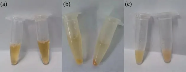
Fig. 1 Sample preparation process
1.2 Western blot
Exosome protein concentrations were measured using a BCA protein assay kit (TaKaRa, China). RIPA lysis buffer (Beyotime,China) was added to lyse the exosomes over 10 min. Then, in the 96-well plates,25 μl of exosome lysate or standard solution is added to each well, solution A with solution B (50∶1) from the kit and added 200 μl was mixed to each well. The specimens are incubated in a drying oven at 37℃ for 30 min. Then, the concentration is measured with an enzyme plate analyzer, and a standard curve is developed. After the concentration is measured, the lysed exosomes are stored at -80℃.
The lysed exosomes were diluted with RIPA lysis buffer, mixed with loading buffer (Beyotime, China),and placed in a water bath at 100℃ for 5-6 min. They were centrifuged and placed on ice. The electrophoresis has been conducted on 10% sodium dodecyl sulfate (SDS) polyacrylamide gel. The proteins were transferred to nitrocellulose membranes.They were blocked at 37℃ for 1 to 2 h with 5% skim milk (0.75 g milk powder + 15 ml phosphate-buffered saline (PBS)). Then the membranes with the primary antibody are incubated at 4℃ overnight(anti-Alix: 1∶1 000; anti-TSG101: 1∶1 000; anti-GAPDH: 1∶10 000; Proteintech, USA). The blot was washed with PBS-T (1 000 ml PBS + 1 ml Tween-20)a total of 3 times, for 10 min each. The blot was then incubated with horseradish peroxidase-conjugated secondary antibody for 1 h at room temperature and visualized using enhanced chemiluminescence reagents.
1.3 Sample processing for AFM
Ultrapure water (200-400 μl) was added to the freshly extracted exosome precipitate (isolation from 1 ml plasma) to resuspend the exosomes. The centrifuge tube was oscillated until the exosomes were dissolved (Figure 1c). The exosome solution is diluted, using ultrapure water, from 80 to 100 times the volume in a new centrifuge tube and the diluted exosome solution is filtered using a 0.22-μm sterileneedle filter. The solution was stored at 4℃ for later use. A nickel-chloride solution is filtered using a 0.22-μm sterile-needle filter. Mica sheets were peeled using adhesive tape to expose a fresh surface. The mica sheets are then placed in a petri dish, and 400 μl nickel-chloride solution is added to their surface for 10 s. The mica sheets is cleaned with ultrapure water,the residual liquid is absorbed with a filter paper, and the mica flakes is allowed to air-dry for 3 min. Then 100 μl of the diluted exosome solution is took and dropped onto the treated mica surface. The petri dish was covered, and incubated at 4℃ overnight. Finally,the mica sheets have been fixed to the AFM testing plate.
As shown in Figure 2a, the AFM imaging experiments were conducted using the commercial Dimension Icon AFM system (Bruker, Santa Barbara,CA) with a ScanAsyst probe made of silicon nitride with a constant cantilever nominal force of 0.7 N/m,an optical-lever sensitivity of 42 nm/V, a tip radius of about 15 nm, and a resonance frequency of about 160 kHz. All AFM experiments were performed under ambient conditions. When the PFT mode was employed, the probe scanned over the specimen surface with a zigzag trajectory. The probe also moved vertically periodically; the force curves were individually recorded. The longitudinal motion frequency of the probe was 2 kHz, and the lateral motion frequency was about 1 Hz. The obtained results, composed of massive force curves, were calculated and converted to multiparametric scanning images of 512×512 pixels.
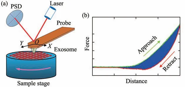
Fig. 2 Schematic of probing living exosomes using the atomic force microscopy-peak force tapping (AFM-PFT)mode
According to the principle of PFT mode, a series of AFM indentation assays was performed on exosomes during scanning experiments. The scanned images consisted of massive force curves, illustrated in Figure 2b. To quantitatively characterize the mechanical properties of exosomes, the force curves were recorded and fitted to the Hertz contact model.The exosomes’ mechanical parameters can be obtained using the formula:
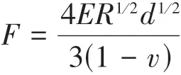
wherevis the Poisson ratio of the exosomes,Ris the radius of pobe,Fis the loading force exerted by the AFM probe,dis the indentation depth, andEis Young’s modulus of the exosomes.
2 Results
2.1 The extracted exosomes express the proteins TSG101 and ALIX
According to relevant literature statistics, one of the methods recognized by researchers for the identification of extracted exosomes is to use Western blot to detect at least two specific marker proteins.The specific marker proteins commonly used are CD63, Tsg101, CD9, CD81, Alix, and HSP70[27-29].Results of Western blot in this experiment showed that the extracted exosomes express the proteins TSG101 and ALIX (Figure 3). Thus, the successful extraction of exosomes was verified.
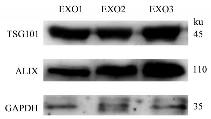
Fig. 3 Validation of exosomes
2.2 Physical characteristics of plasma exosomes based on AFM
A scanned image using the AFM-PFT mode is shown in Figure 4, displaying exosome height information. Figures 4a and 4b show hydrated vesicles of varying sizes without any aggregation or deformation. The height distribution of the exosomes is summarized in Figure 4c. The typical height is ranged from 8 to 10 nm, with a mean value of 8.9 nm and variance of 1.5 nm.
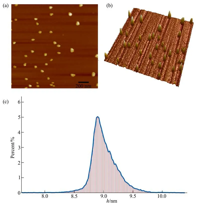
Fig. 4 Morphology calibration of the exosome using AFM-PFT mode
The AFM imaging of exosomes from peripheral blood shows that they are about 15% to 20% of the size of the exosomes obtained from bone marrowbiopsy tissue in our prior study. The morphologic details of single exosome are shown in Figure 5. As demonstrated in Figure 5b, the broadening effect in theX-Yplane caused by the AFM probe tip causes the exosome’s width to appear greater than its height. In fact, the 2 section lines show that the shape of the exosome is nearly spherical. When exosomes are absorbed from the treated mica surface, their height is less than their width. A series of multi-parameter images of exosome specimens is based on the pointby-point force curves obtained using the AFM-PFT mode.
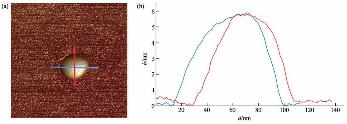
Fig. 5 Exosome height
Figure 6 shows the elastic-stiffness information for an exosome. It should be noticed that elastic stiffness is a relative concept, so the results pictured show the difference in elastic-stiffness values between the mica substrate and the exosome. As shown in Figure 6a, the exosome is softer than mica, and its extent is about 40 MPa, which is calibrated in Figure 6b. We selected dozens of plasma exosomes from lymphoma patients with two different subtypes and found that the stiffness of plasma exosomes from DLBCL patients ((9.45±0.25) MPa) was higher than that from FL patients ((8.23±0.51) MPa) (Figure 6c).Because of its mechanical properties and the deformation data shown in Figure 7a, it can be determined that the exosome is more deformable than the mica substrate. According to the variation curve of exosome deformation, there are more deformations in the interior than at the edge of the exosome(Figure 7b).
Adhesion is another considerable mechanical characteristic; adhesion data are illustrated in Figure 8. The measured adhesion results are evaluated using the energy dissipation process of the force curve. As shown in Figure 2b, energy dissipation can be determined by calculating the area of the blue part encircled by the approach curve and the retrace curve.The dissipation information given in Figure 8 confirms our previous research conclusions[30]. It can be concluded that there is a significant fringe effect at the edge of the exosome, meaning that more energy dissipation is produced there. Figure 8a shows significant energy dissipation around almost the entire edge of the exosome, but previous result[30]shows the phenomenon happening on only a single side of the exosome edge. This important issue should be given more attention in follow-up experiments.
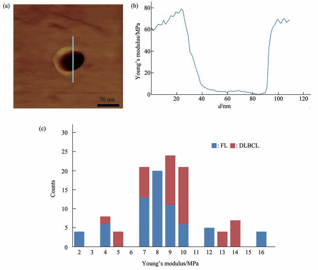
Fig. 6 Elastic stiffness of the exosome in relative Young’s modulus

Fig. 7 Exosome deformation
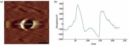
Fig. 8 Adhesion of exosomes in energy dissipation
3 Discussion
The results presented herein are a useful extension and complement to our previous work[26]. In this study, we extracted exosome specimens from the peripheral blood of patients with lymphoma instead of from bone marrow fluid. Exosomes from peripheral blood are more easily available and more clinically significant, as we shall illustrate below.
The traditional method for pathologic tumor diagnosis is to obtain a small amount of tumor tissue through a percutaneous biopsy and to analyze the tumor cellsin vitro. This is an invasive procedure, and biopsy carries certain risks. Sometimes, a pathologic diagnosis cannot be obtained due to the location of the tumor or the physical condition of the patient, limiting the diagnostic and treatment capabilities for the patient. Liquid biopsy technology, as a noninvasive method of diagnosis, can be a complement to tumor biopsy and has been attracting great attention in the diagnosis and treatment of tumors. Exosomes derived from cancer cells enter the bloodstream and act as communication tools in the tumor microenvironment.The genetic information these exosomes carry is highly consistent with that of their parent tumor cells.Studies have shown that tumor cells can produce more exosomes than normal cells, and the amount of exosomes detectable in the peripheral blood of cancer patients is greater than that of healthy subjects[31].Therefore, exosomes in circulating blood are of great significance for “l(fā)iquid biopsy” purposes in patients with cancer. Peripheral blood sampling has the advantages of being convenient and minimally invasive. Blood exosomes can be obtained at any time during tumor treatment; they can also be dynamically monitored as markers of cancer treatment and progression.
Here, the mechanical properties of individual exosomes were analyzed, and it was found that the Young’s modulus of plasma exosomes was different in patients with lymphoma with different aggressiveness. The stiffness of plasma-derived exosomes in patients with highly aggressive lymphoma (DLBCL) was higher than that in patients with inert lymphoma (FL). The correlation between physical properties of exosomes and biological behaviors has also been proposed in previous studies.Yurtseveret al.[32]found in the experiment that the Young’s modulus of exosomes derived from metastatic and invasive tumor cells was increased compared with that of non-metastatic and noninvasive cells. At the same time, they also revealed that this mechanical difference may be related to the enrichment of certain proteins specific to elastic fiber assembly.Further studies on exosome secreting parent cells showed that: the elastic-fiber associated proteins,which were highly expressed in 143B exosomes, were signifificantly less expressed in term parent 143B cells than in nonterm HOS cells. Thus, it is speculated that metastatic tumors may release some proteins through exosomes to maintain the softness of their parent tumor cells. Whiteheadet al.[33]also found that exosomes from non-metastatic cells had lower stiffness than those from metastatic cells, and the adhesion is reduced.
Changes in exosome stiffness of tumor-derived exosomes may be related to the metastatic ability of tumor cells. Some studies have shown that circulating tumor cells (CTCS) can enter circulation through the paracellular pathway of vascular endothelial cells,distant metastasis of the tumor is thus established[34].And exosomes stiffness of malignant and nonmalignant cells is different, this change enhances paracellular transport. Malignant cell-derived exosomes increase the permeability of blood vessels[33,35].
The physical properties of exosomes are not only related to their biological properties, but also,understanding their diameter, length, shape,deformability and stability to mechanical disturbances is also crucial for the development of new methods of exosomes separation (by explosion and microfluidics)and the development of exosomes as drug carriers.
4 Conclusion
The presented research achievements are useful extension and complement for our previous work. The exosome specimens are extracted from the peripheral blood of clinical lymphoma patients instead of bone marrow biopsy. Compared with bone marrow biopsy specimens, peripheral blood specimens are available more easily and more clinically significant. Due to the convenient and less invasive characteristics of peripheral blood, plasma exosomes can be used as biomarkers for dynamic monitoring and analysis of tumor diagnosis and treatment, and the extracted exosomes can be captured by AFM probes through treatment and fixation. Just like our previous AFM scanned results, the exosomes from the peripheral blood show the heterogeneous mechanical properties on the different parts of living exosomes. In addition,differences in physical properties of exosomes from plasma of tumor patients with different pathological subtypes were detected in this study. But some important factors should not be ignored, for example,the exosome size, measured in the AFM experiments,is much smaller than previous ones, which causes smaller deformation under external pressure. The issues will deepen our understanding of the native characteristics of living exosomes from different parts of human body. The continued progress will pave a new road for understanding biomechanics at nanoscale and microscale.