
Repetitive magnetic stimulation affects the microenvironment of nerve regeneration and evoked potentials after spinal cord injury
2016-12-02 07:05:40JinlanJiangXudongGuoShuquanZhangXingangWangShifengWu
中國神經(jīng)再生研究(英文版) 2016年5期
Jin-lan Jiang, Xu-dong Guo, Shu-quan Zhang, Xin-gang Wang, Shi-feng Wu,
1 Scientific Research Center, China-Japan Union Hospital, Jilin University, Changchun, Jilin Province, China
2 Department of Orthopedics, China-Japan Union Hospital, Jilin University, Changchun, Jilin Province, China
3 Department of Cardiovascular Medicine, China-Japan Union Hospital, Jilin University, Changchun, Jilin Province, China
4 Department of Orthopedics, Tianjin Nankai Hospital, Tianjin, China
5 Department of Burns and Plastic Surgery, China-Japan Union Hospital, Jilin University, Changchun, Jilin Province, China
RESEARCH ARTICLE
Repetitive magnetic stimulation affects the microenvironment of nerve regeneration and evoked potentials after spinal cord injury
Jin-lan Jiang1,2,#, Xu-dong Guo3, Shu-quan Zhang4, Xin-gang Wang5, Shi-feng Wu5,*,#
1 Scientific Research Center, China-Japan Union Hospital, Jilin University, Changchun, Jilin Province, China
2 Department of Orthopedics, China-Japan Union Hospital, Jilin University, Changchun, Jilin Province, China
3 Department of Cardiovascular Medicine, China-Japan Union Hospital, Jilin University, Changchun, Jilin Province, China
4 Department of Orthopedics, Tianjin Nankai Hospital, Tianjin, China
5 Department of Burns and Plastic Surgery, China-Japan Union Hospital, Jilin University, Changchun, Jilin Province, China
Graphical Abstract

#These authors contributed equally to this study.
orcid: 0000-0002-7326-3018 (Shi-feng Wu)
Repetitive magnetic stimulation has been shown to alter local blood flow of the brain, excite the corticospinal tract and muscle, and induce motor function recovery. We established a rat model of acute spinal cord injury using the modified Allen's method. After 4 hours of injury, rat models received repetitive magnetic stimulation, with a stimulus intensity of 35% maximum output intensity, 5-Hz frequency, 5 seconds for each sequence, and an interval of 2 minutes. This was repeated for a total of 10 sequences, once a day, 5 days in a week, for 2 consecutive weeks. After repetitive magnetic stimulation, the number of apoptotic cells decreased, matrix metalloproteinase 9/2 gene and protein expression decreased, nestin expression increased, somatosensory and motor-evoked potentials recovered, and motor function recovered in the injured spinal cord. These findings confirm that repetitive magnetic stimulation of the spinal cord improved the microenvironment of neural regeneration, reduced neuronal apoptosis, and induced neuroprotective and repair effects on the injured spinal cord.
nerve regeneration; spinal cord injury; repetitive magnetic stimulation; motor function; rats; rehabilitation; plasticity; regenerative microenvironment; neural regeneration
Introduction
The potential of magnetic stimulation for treating spinal cord injury (SCI) has gradually received greater attention by experts in the field. Magnetic stimulation has been shown to improve cough and respiratory function in SCI patients, improve bowel functions and muscular atrophy of the lower extremities in paraplegic patients, and reduce deep vein thrombosis (Fitch et al., 1999; Hallett, 2007; Paim et al., 2013; Yin et al., 2013). Amar and Levy (1999) verified that microenvironment at the injury site was significantly improved by repetitive magnetic stimulation to the spinal cord, and secondary nerve injury was effectively reduced. Pulses of magnetic stimulation increase blood flow in the capillary bed. Pulse accumulation promotes angiogenesis and indirectly contributes to growth of nerve fibers because of vascular tropism (Crowe et al., 1997; Ohta et al., 2005). Magnetic stimulation reduces Ca2+concentrations, increases Mg2+content, and regulates ion imbalances in the injured spinal cord. Ca2+and Na+-K+-ATPase activities are important regulatory factors for gene expression in neurons(Pommerenke et al., 1996). However, very little is known about how repetitive magnetic stimulation to the spinal cord improves the microenvironment of the injury site and promotes nerve repair, as well as its precise mechanisms of action. The present study explored related indicators of the microenvironment in the injured rat spinal cord using repetitive magnetic stimulation.
Materials and Methods
Ethics statement
This study was approved by the Animal Ethics Committee of China-Japan Union Hospital, Jilin University of China. The animal studies were performed in accordance with the National Institutes of Health Guide for the Care and Use of Laboratory Animals. Precautions were taken to minimize suffering and the number of animals used in each experiment.
Establishment of SCI models
Sixty-seven specific pathogen-free, adult, female, Sprague-Dawley rats, aged 1 month and weighing 250—290 g, were purchased from the Animal Laboratory of Tianjin Medcial University of China (license No. SCXK (Jin) 20070001). The rats were housed at 25°C with 40—60% relative humidity under natural light. Forty-seven rats were intraperitoneally anesthetized with 10% chloral hydrate and fixed on the bench in the prone position. After the lower back was shaved, a median incision was made on the back using the T8—9spinous processes as a center to expose the T7—10spinous processes and the lamina. The T8—9spinous processes and part of the lamina tissue were removed. This exposed dura mater served as the lesion area. Based on the method established by Allen (1940), but with some modifications, a 10-g object was vertically dropped from a 2.5-cm height, which directly impacted the dura mater and T8—9. The wound was washed with hydrogen peroxide, and the tissue was sutured layer by layer. Extrusion was conducted 2—3 times daily to assist with urination until the micturition reflex was recovered. Paralysis of the lower limbs was observed along with tail swinging and spasms. These responses confirmed successful establishment of the model. Model establishment failed in three rats and four rats were excluded because of death. The remaining 40 rats were equally and randomly assigned to the SCI group and repetitive magnetic stimulation group. Twenty rats in the sham surgery group were not exposed to Allen's injury, but the spinal cord was exposed.
Repetitive magnetic stimulation to the spinal cord
Four hours after injury, rats in the repetitive magnetic stimulation group received repetitive magnetic stimulation using a Magstim Rapid2 magnetic stimulator (Magstim, Woburn, MA, USA), with a maximum output intensity of 2.2 T and a 25-mm diameter butterfly coil. The rats received repetitive magnetic stimulation in a supine position and were fixed in a wooden box. The center of the coil was placed at T6—7. Stimulus intensity was 35% of the maximum output intensity. Stimulation was given at a frequency of 5 Hz, with 5 seconds for each sequence, an interval of 2 minutes for 10 sequences, once a day, 5 days in a week, for 2 consecutive weeks.
Evaluation of motor function
Motor functions were assessed before injury, and at 1 and 3 days, and 1, 2, 3, and 4 weeks after injury in the three groups using the modified Tarlov scoring system, the Basso-Beattie-Bresnahan (BBB) locomotor rating scale, and the inclined plane test.
The inclined plane test: rats were horizontally placed on a smooth tiltboard with their heads to the front. The angle was increased every 5°, and the maximum angle at which the rats stayed on the board for 5 seconds was recorded (Wang et al., 2013).
BBB locomotor rating scale: the BBB scores ranged from 0 to 21, where 21 = normal and 0 = complete paralysis. The blind method was used (Wang and Zhang, 2015).
Modified Tarlov scoring system: 0, the lower limbs cannot move or bear weight; 1, the lower limbs can move, but cannot bear weight; 2, the lower limbs can move freely or powerfully, but cannot bear weight; 3, the lower limbs can support weight and walk one or two steps; 4, can walk with mild disturbance; 5, normal walking (Wang and Zhang, 2015).
Determination of apoptotic cells
Three days after injury, five rats from each group were anesthetized with chloral hydrate. After the chest was opened, aortic cannulation was conducted through the left ventricle. Tissue was fixed with 4% paraformaldehyde and a 2-cm long segment of the spinal cord was resected with the injury site as the center. The spinal cord was fixed again in paraformaldehyde, embedded in wax, and sliced into 20-μm thick sections. The sections were then dewaxed. Under an inverted microscope (Leica, Tokyo, Japan), the apoptotic cells were quantified in ten 200× fields. The mean value was then calculated.
Reverse transcription-polymerase chain reaction (RTPCR)
Three days after injury, five rats from each group were randomly selected. Spinal cord tissue (50 mg) was obtained from the injury site at T8—9. In accordance with Trizol reagent instructions (Santa Cruz Biotechnology, Santa Cruz, CA, USA), total RNA was extracted from the spinal cord. RNA content was measured with an ultraviolet spectrophotometer (Olympus, Tokyo, Japan). Using the two-step RT-PCR kit (TaKaRa, Dalian, China), mRNA was reverse-transcribed into cDNA, and cDNA was amplified using PCR. Primer sequences were as follows: matrix metalloproteinase (MMP)2 (414 bp): upstream 5′-TTT TTG TGC CCA AAG AAA GG-3′, downstream 5′-ATG TCA GAC AAC CCG AGT CC-3′; MMP9 (379 bp): upstream 5′-GGT TTC TGT CCA GAC CAA GG-3′, downstream 5′-TGC AAG GAT TGT CAT CTG GA-3′; glyceraldehyde-3-phosphate dehydrogenase (GAP-DH) (300 bp): upstream 5′-GAG GAC CAG GTT GTC TCC TG-3′, and downstream 5′-GGA TGG AAT TGT GAG GGA GA-3′. Amplified products were electrophoresed. Integral optical density analysis of electrophoresis results was conducted using a gel image analysis system (Media Cybernetics, WA, USA). The ratio of integral optical density of MMP9/2 to GAPDH was calculated as the relative expression levels of MMP9/2 mRNA.


Figure 1 Effects of repetitive magnetic stimulation of the spinal cord on motor function in rats with SCI.

Figure 2 Effects of repetitive magnetic stimulation on apoptosis in the injured spinal cord of rats (terminal deoxynucleotidyl transferase dUTP nick-end labeling assay, × 200).
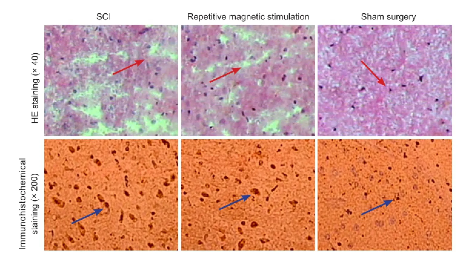
Figure 4 Effects of repetitive magnetic stimulation on repair of the injured spinal cord in rats.

Figure 3 Effects of repetitive magnetic stimulation on MMP 9/2 mRNA (A) and protein (B) expression in the spinal cord of rats with SCI.

Figure 5 Effects of repetitive magnetic stimulation of the spinal cord on nerve conduction in the injured spinal cord of rats.
Western blot assay
Following RNA extraction in RT-PCR, the remaining sample was centrifuged at 1,500 r/min for 30 minutes and the supernatant was collected. The total protein concentration was measured using the Bradford protein assay. Samples were separated by electrophoresis on a 5% stacking gel at 40 V for 1 hour and 10% separating gel at 60 V for 3.5 hours. The separated proteins were transferred onto a membrane using the wet method at 14 V for 14 hours. The membrane was blocked in a swinging bed at 37°C for 2 hours, and washed three times for 10 minutes each. The membranes were incubated with rabbit anti-rat MMP9/2 polyclonal antibody (1:500; Sigma, St. Louis, MO, USA) and rabbit anti-rat GAPDH polyclonal antibody (1:500; Sigma) at room temperature for 60 minutes, followed by three washes with tris-buffered saline/Tween-20 (TBST) for 10 minutes each. The membranes were then incubated with alkaline phosphatase-labeled goat anti-rabbit IgG (1:2,000; Gibco BRL, Gaithersburg, MD, USA) at room temperature for 60 minutes, followed by three washes with TBST for 10 minutes each, followed by tris-buffered saline for 10 minutes. The samples were visualized with 3,3′-diaminobenzidine (Beijing CellChip Biotechnology, Co., Ltd., Beijing, China). Optical density was analyzed using Quantity One analysis software (Hyclone, Logan, UT, USA). The ratio of optical denstiy of MMP9/2 to GAPDH was considered the relative expression level of MMP9/2 protein.
Immunohistochemical staining and hematoxylin-eosin (HE) staining
Four weeks after injury, five rats from each group were anesthetized with 10% chloral hydrate (350 mg/kg) for euthanasia.
Immunohistochemical staining: sections were placed at room temperature for 30 minutes, blocked with fetal bovine serum for 1 hour, washed three times with PBS for 5 minutes each, incubated with mouse anti-rat nestin antibody (1:5,000; Roche) at 4°C overnight, following goat anti-mouse IgG-FITC (1:100) at 37°C for 1 hour, and mounted onto glass slides. The sections were dehydrated through a graded alcohol series, permeabilized with xylene, and mounted with neutral resin. The number of nestin-positive cells was observed in ten 200× fields using an inverted microscope.
HE staining: after anesthesia, the chest was opened to expose the heart. Ascending aortic cannulation was performed. The right auricle was cut open, washed with physiological saline, and fixed with 4% paraformaldehyde. A 1-cm section of spinal cord tissue at the site of injury was collected, dehydrated through a graded alcohol series, and sliced into 20-μm thick longitudinal frozen sections. The sections were then stained with hematoxylin for 5 minutes, washed with running water, differentiated with hydrochloric acid in ethanol for 10 seconds, washed with running water for 10 minutes, stained with eosin for 7 minutes, washed with running water, dehydrated through a graded alcohol series, permeabilized with xylene, and mounted with neutral resin.
Detection of somatosensory-evoked potential (SEP) and motor-evoked potential (MEP)
Four weeks after injury, five rats each from the sham surgery group, SCI group, and repetitive magnetic stimulation group were intraperitoneally anesthetized with 10% chloral hydrate (350 mg/kg). SEP and MEP were measured using a Keypoint 4-evoked Potential System (Beijing Weidi Kangtai Medical Instrument Co., Ltd., Beijing, China).
SEP: The rats were placed in a horizontal plane. The hind limbs were stimulated with a stimulating electrode. A recording electrode was placed at the intersection of the coronal suture and sagittal suture healing line under the scalp (i.e., hindlimb cortical sensory area). A reference electrode was placed 0.5 cm posterior to the hindlimb cortical sensory area. A direct current, square wave, and electrical pulses were given until the hind limb exhibited a slight tic. The conditions were as follows: current intensity of 5—15 mA, pulse width of 0.2 ms, frequency of 3 Hz, superimposed: 50—60 times. SEP latency and amplitude were recorded.
MEP: The stimulating electrodes were placed 2 mm anterior to the coronal suture and 2 mm lateral to the sagittal suture under the scalp (i.e., motor cortex). The electrodes were stimulated as follows: stimulus intensity of 40 mA, pulse width of 0.1 ms, frequency of 1 Hz, sensitivity of 5 μV/ D, scanning speed of 5 ms/D; superimposed: 300—500 times. MEP latency and amplitude were recorded.
Statistical analysis
All data were expressed as the mean ± SD, and analyzed with SPSS 17.0 software (SPSS, Chicago, IL, USA). Continuous variable among groups was compared using analysis of variance for randomized block design. Continuous variable between groups was compared using paired t-test. An alpha of 0.05 with a two-tailed test was used.
Results
Repetitive magnetic stimulation improved motor funcion in SCI rats
There was no significant difference in BBB locomotor rating scale scores, inclined plane test scores, or modified Tarlov scoring system scores in all rats prior to model establishment. BBB locomotor rating scale scores, inclined plane test scores, and modified Tarlov scoring system scores were significantly greater in the repetitive magnetic stimulation group than in the SCI group (2—4 weeks after injury; P < 0.05). The above scores were significantly less in the SCI and repetitive magnetic stimulation groupsthan in the sham surgery group at 2—4 weeks after injury (P< 0.05; Figure 1).
Repetitive magnetic stimulation of the spinal cord reduced apoptosis in the injured spinal cord
TUNEL assay results revealed significantly less apoptotic cells in the repetitive magnetic stimulation group (13.67 ± 2.32/200× field) than in the SCI group (32.76 ± 3.44/200× field) (P < 0.05). No apoptotic cells were identified in the sham surgery group (Figure 2).
Repetitive magnetic stimulation of the spinal cord reduced MMP9/2 mRNA and protein expression in the injured spinal cord
RT-PCR and western blot assay results demonstrated significantly increased MMP9/2 mRNA and protein expression at 72 hours after injury (P < 0.01). MMP9/2 mRNA and protein expression in the repetitive magnetic stimulation group was less than in the SCI group (P < 0.05), but more than in the sham surgery group (P < 0.05; Figure 3).
Repetitive magnetic stimulation of the spinal cord improved pathomorphology and increased the number of nestin-positive cells in the injured spinal cord
Four weeks after injury, hematoxylin-eosin and immunohistochemical staining revealed a complete and clear structure spinal cord, with no cavity and densely arranged nerve fibers, in the sham surgery group. A large number of nestin-positive cells were quantified (30.42 ± 3.83/200× field). In the SCI group, the spinal cord tissue exhibited a loose structure, with a visible cavity and a large number of necrotic neurons. The nerve fibers were loosely arranged and appeared shorter and less compared with the repetitive magnetic stimulation group. There were fewer nestin-positive cells in the SCI group (5.83 ± 1.72/200× field) than in the sham surgery group (P < 0.05). In the repetitive magnetic stimulation group, loose spinal cord tissue and a small cavity were visible, with a recovered density of nerve fibers. The number of nestin-positive cells was slightly higher in the repetitive magnetic stimulation group (19.24 ± 2.20/200× field) than in the SCI group (P < 0.05; Figure 4).
Repetitive magnetic stimulation of the spinal cord improved electrophysiological function in the injured spinal cord
After model establishment, the evoked potential waveform disappeared in the repetitive magnetic stimulation and SCI groups. Four weeks later, SEP and MEP were slightly recovered in the SCI group compared with the sham surgery group (P < 0.05). SEP and MEP were significantly recovered, latencies were shorter, and amplitudes were higher in the repetitive magnetic stimulation group than in the SCI group (P< 0.05; Figure 5).
Discussion
Repetitive magnetic stimulation of the spinal cord can reverse synaptic function at the site of injury, improve neuronal plasticity, and protect neurons against external factor-induced degeneration and necrosis. Long-term repetitive magnetic stimulation can increase mRNA expression of brain-derived neurotrophic factor (Muller et al., 2000) and reduce c-fos protein levels at the injury site (Hausmann et al., 2000), which suggests a neuroprotective effect of repetitive magnetic stimulation of the spinal cord. Consequently, the neuroprotective effect of repetitive magnetic stimulation has been shown to indirectly reduce proliferation of reactive astrocytes, as well as decrease axonal sprouting, synaptic reconstruction, and excitatory loop formation (Avoli, 1996; Muller et al., 2000; Kudo et al., 2005).
Following central nervous system injury, MMP2 and MMP9 disrupt the tight connections between capillaries, basement membranes, and the blood brain barrier, and induce vasogenic edema of the central nervous system (Liu and Shubayev, 2011; Yang et al., 2013; Lee et al., 2014). Results from the present study suggested that repetitive magnetic stimulation effectively reduced edema-related gene and protein expression at the injury site, which could also be responsible for reducing the degree of edema. Repetitive magnetic stimulation of the spinal cord also effectively reduced neuronal apoptosis at the injury site. Results from the BBB locomotor rating scale, inclined plane test, and modified Tarlov scoring system showed that repetitive magnetic stimulation of the spinal cord improved motor function of the hind limbs in rats. Repetitive magnetic stimulation shortened SEP and MEP latencies and increased amplitudes in SCI rats, and effectively contributed to functional recovery of the injured spinal cord. This study provides a new theoretical basis for repetitive magnetic stimulation in SCI repair.
Author contributions: SFW and JLJ conceived and designed the study, provided data and ensured the integrity of the data. SQZ analyzed the data, wrote the paper, obtained the funding, provided technical or material support and served as a principle investigator. XDG and XGW were in charge of paper authorization and statistical analysis. All authors approved the final version of the paper.
Conflicts of interest: None declared.
Plagiarism check: This paper was screened twice using Cross-Check to verify originality before publication.
Peer review: This paper was double-blinded and stringently reviewed by international expert reviewers.
Allen IM (1940) The immediate and remote effects of minor lesions of the cervical portion of the spinal cord following head injury. Aust N Z J Surg 10:157-172.
Amar AP, Levy ML (1999) Pathogenesis and pharmacological strategies for mitigating secondary damage in acute spinal cord injury. Neurosurgery 44:1027-1039.
Avoli M (1996) GABA-mediated synchronous potentials and seizure generation. Epilepsia 37:1035-1042.
Crowe MJ, Bresnahan JC, Shuman SL, Masters JN, Beattie MS (1997) Apoptosis and delayed degeneration after spinal cord injury in rats and monkeys. Nat Med 3:73-76.
Fitch MT, Doller C, Combs CK, Landreth GE, Silver J (1999) Cellular and molecular mechanisms of glial scarring and progressive cavitation: in vivo and in vitro analysis of inflammation-induced secondary injury after CNS trauma. J Neurosci 19:8182-8198.
Hallett M (2007) Transcranial magnetic stimulation: a primer. Neuron 55:187-199.
Hausmann A, Weis C, Marksteiner J, Hinterhuber H, Humpel C (2000) Chronic repetitive transcranial magnetic stimulation enhances c-fos in the parietal cortex and hippocampus. Mol Brain Res 76:355-362.
Kudo K, Yamada M, Takahashi K, Nishioka G, Tanaka S, Hashiguchi T, Fukuzako H, Takigawa M, Higuchi T, Momose K, Kamijima K, Yamada M (2005) Repetitive transcranial magnetic stimulation induces kf-1 expression in the rat brain. Life Sci 76:2421-2429.
Lee JY, Choi HY, Na WH, Ju BG, Yune TY (2014) Ghrelin inhibits BSCB disruption/hemorrhage by attenuating MMP-9 and SUR1/TrpM4 expression and activation after spinal cord injury. Biochim Biophys Acta 1842:2403-2412.
Liu H, Shubayev V (2011) Matrix metalloproteinase-9 controls proliferation of NG2+progenitor cells immediately after spinal cord injury. Exp Neurol 231:236-246.
Muller MB, Toschi N, Kresse AE, Post A, Keck ME (2000) Long-term repetitive transcranial magnetic stimulation increases the expression of brain-derived neurotrophic factor and cholecystokinin mRNA, but not neuropeptide tyrosine mRNA in specific areas of rat brain. Neuropsychopharmacology 23:205-215.
Ohta S, Iwashita Y, Takada H, Kuno S, Nakamura T (2005) Neuroprotection and enhanced recovery with edaravone after acute spinal cord injury in rats. Spine 30:1154-1158.
Paim LR, Schreiber R, Matos-Souza JR, Silva AA, Campos LF, Azevedo ER, Alonso K, de Rossi G, Etchebehere M, Gorla JI, Cliquet A Jr, Nadruz W Jr (2013) Oxidized low-density lipoprotein, matrix-metalloproteinase-8 and carotid atherosclerosis in spinal cord injured subjects. Atherosclerosis 231:341-345.
Pommerenke H, Schreiber E, Dürr F, Nebe B, Hahnel C, M?ller W, Rychly J (1996) Stimulation of integrin receptors using a magnetic drag force device induces an intracellular free calcium response. Eur J Cell Biol 70:157-164.
Wang D, Zhang J (2015) Effects of hypothermia combined with neural stem cell transplantation on recovery of neurological function in rats with spinal cord injury. Mol Med Report 11:1759-1767.
Wang D, Fan YH, Zhang JJ (2013) Transplantation of Nogo-66 receptor gene-silenced cells in a poly(D,L-lactic-co-glycolic acid) scaffold for the treatment of spinal cord injury. Neural Regen Res 8:677-685.
Yang J, Wang G, Gao C, Shao G, Kang N (2013) Effects of hyperbaric oxygen on MMP-2 and MMP-9 expression and spinal cord edema after spinal cord injury. Life Sci 93:1033-1038.
Yin Y, Sun W, Li Z, Zhang B, Cui H, Deng L, Xie P, Xiang J, Zou J (2013) Effects of combining methylprednisolone with rolipram on functional recovery in adult rats following spinal cord injury. Neurochem Int 62:903-912.
Copyedited by Cooper C, Frenchman B, Yu J, Qiu Y, Li CH, Song LP, Zhao M
NEWS Congratulation to Professor Kowk-fai So of him being named as a Fellow of the National Academy of Inventors doi: 10.4103/1673-5374.182716 Professor Kwok-fai So, Editor-in-Chief of Neural Regeneration Research, has been named a Fellow of the National Academy of Inventors (NAI). 168 NAI Fellows were inducted on April 15, 2016, as part of the Fifth Annual Conference of the National Academy of Inventors at the USPTO in Alexandria, Virginia.
Professor Kwok-fai So currently serves as Director of GHM Institute of Neural Regeneration, Jinan University and Jessie Ho Professor in Neuroscience, Department of Ophthalmology of Li Ka Shing Faculty of Medicine, The University of Hong Kong. Professor So is one of the pioneers in the field of axonal regeneration in visual system, and he is a leader in the area of neuroprotection and neuroregeneration of the mammalian optic nerve, spinal cord and brain. He is Co-Chairman of the Board of Director of the China Spinal Cord Injury Network and Director of Hong Kong Spinal Cord Fund (HKSCIFund). He is the recipient of China National Natural Science Award, China Spinal Cord Injury Rehabilitation Contribution Award, Rick Hansen Difference Maker Award, Everfront Award for Stem Cells Research. He has published 303 articles, two books and 70 review articles. Other than serving as Editor-in-Chief of Neural Regeneration Research, Professor So is also member of editorial board of 14 journals, including Cell Transplantation, Restorative Neurology and Neuroscience, Frontiers in Aging Neuroscience and Chinese Journal of Neuroscience. He is a member of the Chinese Academy of Sciences as well.
Professor So holds 4 U.S. patents, 16 international patents and 3 China patents, with 16 patents in the field of hemostasis (stop bleeding) and sealant (control leaking). His most significant patents are Compositions and Methods for Promoting Hemostasis and Other Physiological Activities and Compositions and Methods for Affecting Movement of Contaminants, Bodily Fluids or Other Entities, and/or Affecting Other Physiological Conditions, and they have been licensed to Arch Therapeutics. These patents describe compositions that include nanoscale-structured materials or precursors thereof (e.g., self-assembling peptides), as well as methods for using the compositions to promote hemostasis, to protect the skin or wounds from contamination. It is significant because it can be developed into a new product that can seal and protect leaking and bleeding tissue. This may transform the landscape of interventional healthcare.
Founded in 2010, the National Academy of Inventors is a non-profit member organization comprised of U.S. and international universities, and governmental and non-profit research institutions, with over 3,000 individual inventor members and fellows spanning more than 200 institutions, and growing rapidly. It was to recognize and encourage inventors with patents issued from the U.S. Patent and Trademark Office, enhance the visibility of academic technology and innovation, encourage the disclosure of intellectual property, educate and mentor innovative students, and translate the inventions of its members to benefit society.
10.4103/1673-5374.182710 http://www.nrronline.org/
How to cite this article: Jiang JL, Guo XD, Zhang SQ, Wang XG, Wu SF (2016) Repetitive magnetic stimulation affects the microenvironment of nerve regeneration and evoked potentials after spinal cord injury. Neural Regen Res 11(5)∶816-822.
Accepted: 2016-01-26
*Correspondence to: Shi-feng Wu, wsf19770620@126.com.
- 中國神經(jīng)再生研究(英文版)的其它文章
- Recovery of injured fornical crura following neurosurgical operation of a brain tumor: a case report
- Gender difference in the neuroprotective effect of rat bone marrow mesenchymal cells against hypoxiainduced apoptosis of retinal ganglion cells
- Vitamin B complex and vitamin B12levels after peripheral nerve injury
- Methylprednisolone microsphere sustained-release membrane inhibits scar formation at the site of peripheral nerve lesion
- A self-made, low-cost infrared system for evaluating the sciatic functional index in mice
- Methylprednisolone exerts neuroprotective effects by regulating autophagy and apoptosis