The Role of Clinical Cardiac Magnetic Resonance Imaging in China: Current Status and the Future
Shi Chen, MD, Qing Zhang, MD and Yucheng Chen, MD
1Cardiology Division, West China Hospital, Sichuan University, Chengdu, Sichuan 610041, China
Introduction
As a powerful noninvasive cardiovascular imaging modality, cardiac magnetic resonance (CMR) imaging plays an important role in the diagnosis and management of cardiovascular diseases. CMR imaging has many advantages in cardiac imaging and is considered a “one-stop shop” to provide comprehensive information on the heart. Recently, dedicated guidelines or consensus on CMR scanning, postprocessing, reporting, and clinical appropriateness [1–4]have been published, providing standardized guidance for the clinical utility of CMR imaging. CMR imaging is emerging in Chinese hospitals and has a promising future. This article will brie fly describe the clinical application of CMR imaging in China and discuss obstacles for its future development.
History and Current Status of Clinical CMR Imaging in China
The clinical application of CMR imaging was initiated in the late 1980s [5, 6], when the clinical feasibility of CMR imaging was validated to show cardiac structure. In the late 1990s, the clinical role of CMR imaging became important because of the use of steady-state free precession sequence and LGE. In China, the earliest application of CMR imaging in patients was in Nanfang Hospital, Guangzhou, in the 1980s. Then several reports of CMR studies were published at the end of the 1980s and in the early 1990s [5–7]. However, real-word practice of CMR imaging in the clinical setting in China was very rare,especially in the late 1990s to the early 2000s, when there were very sporadic clinical reports. Clinical CMR imaging was available in only a very limited number of hospitals; for example Fuwai Hospital[8, 9]. However, the situation has begun to change gradually in the past decade; more and more physicians in radiology and cardiology departments have been promoting the use of CMR imaging in clinical practice. On the basis of a brief survey conducted in 2015 to investigate current CMR imaging practice in mainland China, 77 of the 108 participating hospitals reported that they routinely perform clinical CMR imaging. Most of them (63, 82%) were university or academic hospitals. About half of these hospitals performed 50–200 scans per year and only five centers performed more than 500 scans per year. For example, the number of clinical CMR scans in West China Hospital increased from 20 per year to more than 1000 per year from 2009 to 2015, but this still lags far behind clinical demands. The common reasons for CMR imaging are suspected cardiomyopathies,mostly nonischemic, including hypertrophic cardiomyopathy (HCM; 24.6%), dilated cardiomyopathy(DCM; 17.9%), amyloidosis (2.5%), restrictive cardiomyopathy (2.1%), arrhythmogenic right ventricular cardiomyopathy (ARVC; 1.1%), left ventricular noncompaction (0.4%), and Fabry disease (0.4%);only 21.1% of patients were referred for the evaluation of viability after myocardial infarction [10].The major referral reasons are similar among highvolume centers. In addition, the survey showed that there are state-of-the-art scanners in most hospitals,which provide a good means to promote the clinical utility of CMR imaging.
Clinical Role of CMR Imaging in China
CMR Imaging in Ischemic Heart Disease
Ischemic heart disease is a major cardiovascular disease in China. There are 2 million patients with myocardial infarction and almost 500,000 new cases of myocardial infarction annually [11]. CMR imaging has robust ability in the diagnosis and risk strati fication of ischemic heart disease by perfusion,viability, and angiography.
Stress CMR imaging has been used to detect ischemia in coronary artery disease, and has been demonstrated to be better than myocardial perfusion single photon emission computed tomography.However, the extra needs of stress-inducing medication, continuous monitoring devices, and on-site physicians can restrict its routine clinical application in China. From a search for “stress CMR” and “China”in all available databases, including PubMed, Ovid,and Chinese Wanfang databases, there are very few reports on stress CMR imaging performed in patients with suspected ischemic heart disease. Pan et al. [12]performed a clinical study in 71 patients using quantitative stress perfusion CMR imaging and found that myocardial transmural perfusion gradient is a better parameter than conventional transmural perfusion analysis to detect myocardial ischemia when compared with fractional flow reserve.
In 2000 Kim et al. [13] reported a landmark study that showed the transmural extent of infarct assessment by late gadolinium enhancement (LGE)could be a novel parameter for myocardial viability.Potentially, viability evaluation by means of LGE could be used to guide revascularization in ischemic heart disease. In China, Li et al. [14] evaluated the effect of percutaneous coronary intervention on left ventricular function in patients with myocardial infarction assessed by LGE and found results similar to those of Kim et al. Additional studies confirmed that the ability of CMR imaging to identify viable myocardium was not inferior to that of single photon emission computed tomography [15, 16].The health care cost for cardiovascular diseases has largely increased in recent years, with a total hospitalization expenditure of 40 billion yuan, which is estimated to be 1.64% of the national health expenditures [17]. Although it helps to improve the cost-effectiveness of myocardial revascularization,myocardial viability evaluation by CMR imaging is still underused because to its low availability in China. In addition to LGE, recent studies have revealed that the extracellular volume (ECV) fraction may serve as a promising alternative parameter in assessing regional myocardial injury that could predict recovery after revascularization. Because of its quantitative nature, ECV fraction would be highly reliable and reproducible. Chen et al. [18]reported in a small cohort of patients with chronic total coronary occlusion that the ECV fraction provided a measure additional to delayed enhancement imaging in assessing functional recovery after revascularization. They proposed a novel risk stratification model, including myocardial necrosis and functional abnormalities derived from CMR imaging in ST segment elevation myocardial infarction patients, that could predict the major cardiovascular adverse events at 90-day follow-up [19].
Coronary magnetic resonance angiography(CMRA) was first introduced in 1987 [20]. CMRA shows good accuracy in the diagnosis of coronary stenosis with the development of imaging techniques.It remains a component of the imaging protocol for ischemic heart disease, especially for patients allergic to conventional contrast media. Some small-scale clinical trials in China have demonstrated that contrast-enhanced whole-heart CMRA using a spoiled gradient echo sequence at 3.0 T provided imaging with superior accuracy, relatively high spatial resolution, and a short imaging time. By using this method,Yang et al. [21] demonstrated the feasibility of accurate detection of significant stenosis in coronary arterial segments larger than 1.5 mm. Chen et al. [22]suggested that image quality can be further improved by sublingual nitroglycerin and abdominal banding.One study that combined CMRA with 3D volumetargeted thin-slab fast imaging employing steadystate acquisition (FIESTA) found a similar accuracy in detecting proximal coronary stenosis between CMRA and computed tomograph angiography [23].Jin et al. [24] compared volume-targeted acquisition with whole-heart acquisition in 1.5-T free-breathing 3D CMRA and found that right coronary artery/left circum flex artery–targeted and left coronary artery/left anterior descending coronary artery–targeted acquisition yielded higher vessel sharpness and overall image quality [24]. The whole-heart method was superior to the volume-targeted method in the visualization of the posterior descending artery. Therefore the different methods of acquiring CMRA images are not competing imaging approaches but have different indications for clinical use. It has been reported that coronary anatomy, myocardial damage, and viability information can be feasibly acquired in one CMR examination in some Chinese heart centers [25].
CMR Imaging in Nonischemic Cardiomyopathy
Cardiomyopathy is a heterogeneous group of diseases. CMR imaging has been established as a robust technique in the evaluation of cardiomyopathies.CMR imaging not only provides comprehensive information on structure and function, but also performs tissue characterization to establish the cause of cardiomyopathy. Not surprisingly, further investigation on nonischemic cardiomyopathies is the most common referral reason for CMR imaging in China.
Dilated Cardiomyopathy
Dilated cardiomyopathy (DCM) is a common cause of heart failure and is characterized by a dilated left ventricle with impaired systolic function in the absence of coronary artery disease and other nonmyocardial causes of dilatation. Left ventricular volumes and left ventricular ejection fraction provide fundamental measures of function and serve as strong prognostic indicators for patients with DCM. CMR imaging is the gold standard for the measurement of ventricular function. More importantly, LGE-CMR imaging, as a gold standard for myocardial scar, is widely accepted in the differential diagnosis of DCM as well as for outcome prediction. In DCM patients, a midline scar, especially in the interventricular septum, is a specific presentation in CMR imaging (Figure 1). In a DCM cohort study, Li et al. [26] repotted that DCM patients with diffuse LGE had higher mortality than patients with LGE in the interventricular septum or LGE in a location other than the interventricular septum. In another prospective study, Lu et al. [27] found that fat deposition is a common phenomenon in DCM, and is associated with fibrosis volume and left ventricular function.However, the cause and prognostic significance need to be further explored. Studies have compared CMR features of isolated left ventricular noncompaction and DCM in Chinese adults and demonstrated some morphological and functional differences; for example, a higher end-diastolic noncompaction/compaction ratio in patients with left ventricular noncompaction [28, 29].
Hypertrophic Cardiomyopathy
Hypertrophic cardiomyopathy (HCM) is a genetic cardiac disease characterized by various degrees of myocardial hypertrophy. The estimated prevalence of HCM in China is 0.13% [30], and therefore at least 1 million patients have this disease. Because of high-quality imaging (Figure 2), CMR imaging was used to investigate HCM patients in China more than 2 decades ago.
CMR imaging can clearly depict the morphology of the septum and left ventricular out flow tract(LVOT). The hypertrophic patterns of the septum are critical determinants of blood flow and function, but are variable. Zhong et al. [31] developed an independent coordinate method to quantify the regional shape of the left ventricle in terms of curvature by CMR imaging to identify the morphology subtype of HCM. Furthermore, Yan et al. [32]focused on the long-term outcome of HCM patients with mid-ventricular obstruction as a rare form of HCM. The results indicated that in Chinese patients,mid-ventricular obstructive HCM is associated with an unfavorable prognosis in terms of cardiovascular death. Additional studies investigated ventricular function in Chinese HCM patients by means of novel parameters and found that longitudinal strain was commonly impaired [33] and that right ventricular systolic function was associated with symptomatic severity [34].
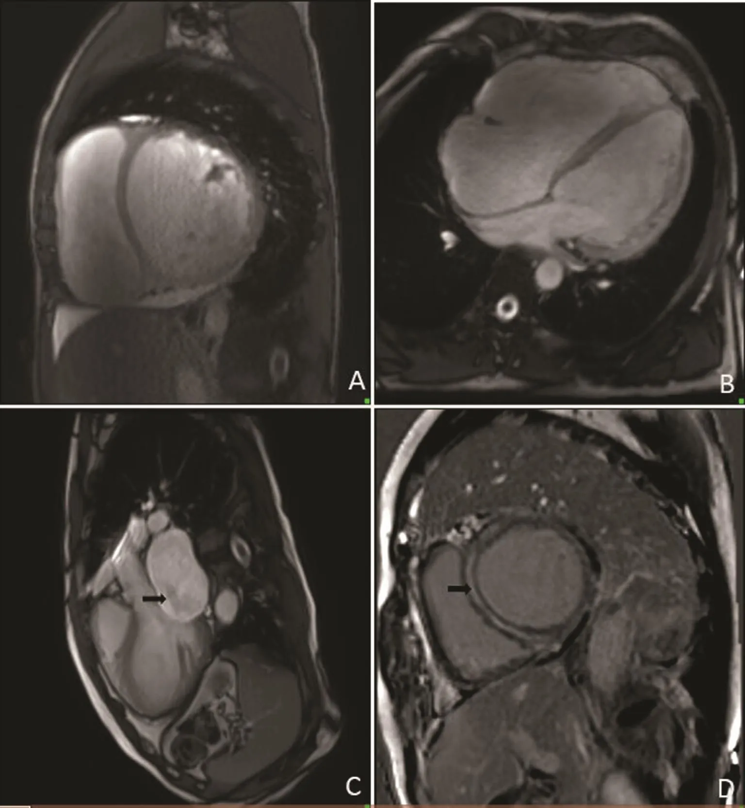
Figure 1 The CMR Images of DCM.A and B are short-axis and 4 chamber view of cine image showing dilated cardiac chambers, respectively. Additionally, A shows pericardial effusion. C is 3 chamber view of cine image which shows systolic mitral regurgitation (black arrow). D is short-axis of LGE image which shows midline hyperenhancement on LGE in anterior septum, septum and inferior wall (black arrow).
Myocardial fibrosis is a very important pathologic characteristic of HCM, and can be observed with LGE-CMR imaging. LGE at the anterior and inferior insertions of the right ventricle is the most typical replacement fibrosis pattern. Apical HCM(ApHCM) is a special subtype of HCM for which there are more patients in the Far East. Guo et al.[35] observed a special pattern of LGE distribution in Chinese ApHCM patients. LGE was found not only in the thickened apex but also in other left ventricular segments irrespective of the presence or absence of hypertrophy. Studies have demonstrated that LGE is an independent risk factor for sudden death, heart failure progression, and ventricular arrhythmia in HCM patients, which was also revealed in Chinese patients [36]. Nevertheless,this relationship has not been con firmed in Chinese ApHCM patients [35].
Percutaneous transluminal septal myocardial ablation (PTSMA) as an alternative to surgical myectomy to relieve LVOT obstruction was first reported in 1995 [37]. Thousands of HCM patients have received this treatment in China. Lu et al. [38]reported good correlations between the reduction of postoperative LVOT gradient and the thickness of the basal anterior segment, the thickness of the basal anteroseptal segment, and the total thickness of these two segments. Moreover, they investigated and supported the value of the use of CMR imaging during patient selection for the PTSMA procedure. Yuan et al. [39] demonstrated the feasibility of CMR imaging in the short-term and long-term follow-up after PTSMA. Another 16-month follow-up study involving CMR imaging revealed that PTSMA not only significantly reduced LVOT obstruction but also reversed biventricular remodeling in terms of decreased right ventricular and left ventricular masses and increased right ventricular and left ventricular end-diastolic volumes [40]. In addition, significant and sustained improvement in left ventricular diastolic function has been observed after PTSMA [41].
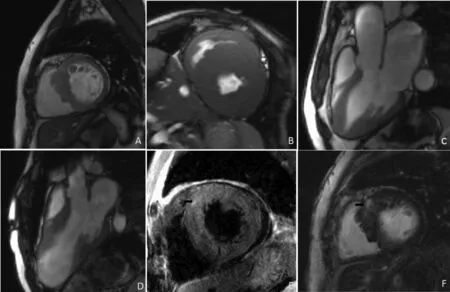
Figure 2 The CMR Images of HCM.Both A and B are short-axis view of cine images. A demonstrates asymmetric hypertrophy of the septum. B shows homogenous hypertrophy of LV and RV is also involved. Cine images in 3 chambers view (C and D) show some special phenotypes of HCM including apical hypertrophy and mid left ventricular hypertrophy with thinning myocardium and aneurysm at the apical segments. T2-weighted image in short axis view (E) reveals high signal area which indicates myocardial edema in the anterior insertions of the RV (black arrow). LGE short-axis view (F) the anterior insertions of the RV (black arrow).
Arrhythmogenic Right Ventricular Cardiomyopathy
Arrhythmogenic right ventricular cardiomyopathy (ARVC) is an inherited disease that primarily affects the right ventricle. The clinical presentation is usually malignant arrhythmia or sudden cardiac death. A 2010 ARVC task force de fined right ventricular kinesis and right ventricular ejection fraction measured by CMR imaging as major criteria for the diagnosis of ARVC [42], in which CMR imaging was strongly recommended as the optimal imaging modality to significantly increase the sensitivity of detection (Figure 3). However,only two small cohort studies have validated the feasibility of CMR imaging for the diagnosis of ARVC in Chinese patients [43–45]. The prominent clinical pro file of Chinese patients with ARVC includes relatively low incidence of premature sudden death and uncommon left ventricular involvement [43]. Cheng et al. [46] reported that N-terminal prohormone of brain natriuretic peptide was associated with the extent of right ventricular dilatation and dysfunction measured by CMR imaging in patients with ARVC, and may be considered a prognostic marker. In addition, Ma et al. [47] found that right ventricular out flow tract area, right ventricular end-diastolic volume, and end-systolic volume measured by CMR imaging were positively correlated to the extent of QRS dispersion on a Holter recording. In a cohort study that included 193 Chinese patients with ARVC,10 intracardiac thrombi were identi fied in eight patients; most of them (7 of 10) were found in the right ventricular apex [48]. Multivariate analysis showed that female sex and left ventricular dysfunction are independently associated with a high risk of ventricular thrombosis in ARVC.
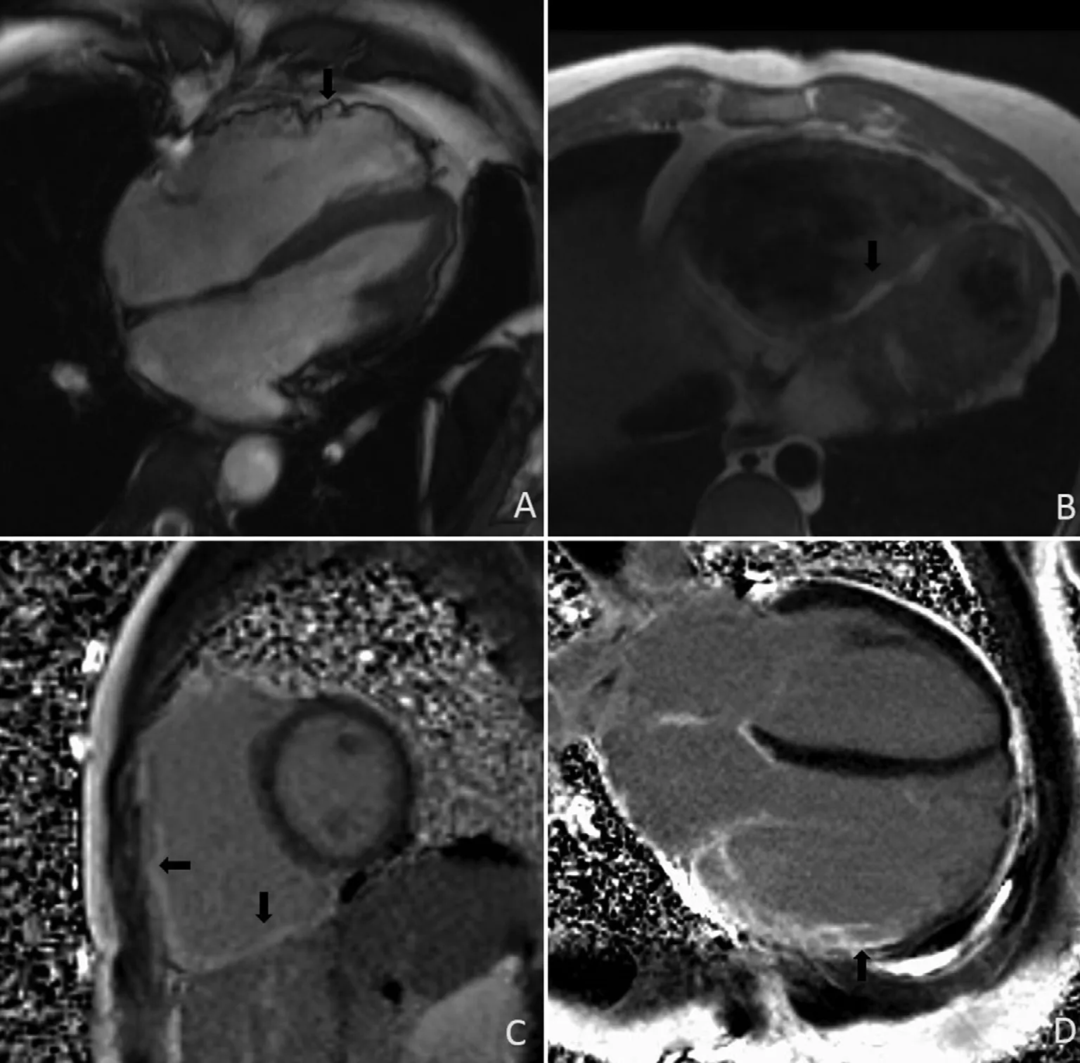
Figure 3 The CMR Images of ARVC.Four-chamber cine image (A) shows marked enlargement of the RV, impairment of global RV function, segmental minimal aneurysm of the RV wall (black arrow). Short-axial T1-weighted fast spin-echo image (B) detects fatty in filtration of RV (black arrow). Both C and D are LGE images with PSIR which demonstrate the hyperenhancement of RV (black arrow).
CMR Imaging in Myocarditis
Myocarditis is an in flammatory myocardial disease that may be caused by different pathogens and triggers. The diagnosis of myocarditis remains a difficult clinical scenario, since the clinical spectrum of myocarditis extends from mild or no symptoms to acute cardiogenic shock. CMR imaging is currently the best noninvasive imaging modality to con firm the diagnosis of myocarditis,with its ability to detect focal in flammation and scarring (Figure 4). In the United States and other Western countries, CMR imaging is commonly performed in patients presenting with acute chest pain and raised levels of myocardial injury biomarkers after normal coronary arteries are demonstrated. However, CMR imaging is not yet a routine imaging modality in patients suspected of having myocarditis, probably because of the long waiting time and the high cost of the examination.In a cohort study that enrolled only 30 patients with suspected myocarditis, the diagnostic values of different CMR scan methods (edema imaging,global relative enhancement, LGE) involved in the Lake Louise criteria [49] were compared. The results recon firmed that CMR imaging is an excellent imaging modality. Liu et al. [50] described the variety of CMR findings in children with myocarditis, including regional or global myocardial signal increase in T2-weighted images and early gadolinium enhancement and LGE in T1-weighted images. In addition, some complicated cases of myocarditis detected by CMR imaging were reported [51, 52].
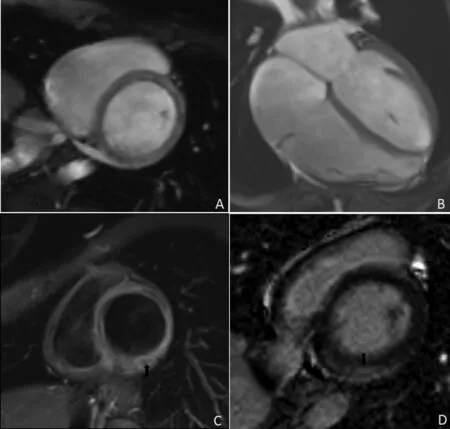
Figure 4 The CMR Images of Myocarditis.Both the short-axis (A) and the four-chamber (B) cine image show the mildly enlargement of cardiac chambers. Moreover,pericardial effusion can also be detected in myocarditis (B). T2-weighted image (C) show high signal which indicate myocardial edema in inferolateral region (black arrow). LGE image (D) show subepicardial layer of hyperenhancement in inferior wall of LV (black arrow).
CMR Imaging in Congenital Heart Disease
CMR imaging is an ideal test for the initial diagnosis and follow-up of congenital heart disease because it is noninvasive and does not expose patients to ionizing radiation. Recently, we demonstrated by using CMR imaging in a group of patients with Ebstein anomaly that cone reconstruction as a novel surgical procedure for tricuspid valve repair yielded a good short-term survival [53]. Cone reconstruction reduced functional right ventricular volume, improved right ventricular global synchronization, and restored right ventricular geometry. Contrast-enhanced magnetic resonance angiography (MRA) is a noninvasive modality for unlimited visualization of the aortic arch; however,only a few studies have demonstrated the feasibility of this imaging modality for the detection of congenital aortic arch anomalies in Chinese patients [54, 55].Safety issues related to gadolinium-based contrast agents, in particular those with reduced renal function, are major concerns. Therefore Chang et al. [56]adopted a novel unenhanced CMR technique based on 3D steady-state free precession to compare it with contrast-enhanced MRA. This novel technique provides powerful complementary information for patients who undergo contrast-enhanced MRA, especially at the ventriculoarterial connection site.
New CMR Techniques for Clinic al Research in China
In recent years, quantitative CMR techniques, such as T1, T2, and T2* mapping, have shown their potential unique clinical and research values. Gao et al. [57]reported that the native myocardial T1 values were significantly increased in hypothyroidism patients compared with healthy controls and that increased T1 values were correlated with free triiodothyronine concentration and cardiac function impairment. The study indicated that diffuse myocardial fibrosis was common in hypothyroidism patients. Myocardial ECV fraction is de fined as the percentage of extracellular space, which is measured by use of myocardial T1 imaging before and after contrast agent administration, and calibrated with hematocrit. Chen et al.[18] recently demonstrated that ECV fraction was able to assess myocardial injury and angiographic collateral flow in patients with chronic total coronary occlusion. In addition, ECV fraction could predict functional recovery after revascularization in these patients [18]. The T2 value re flects the content of water in the myocardium, which is considered a sensitive parameter to detect myocardial edema, a common pathophysiologic feature in some in flammatory myocardial diseases or acute myocardial injury. T2 mapping techniques as a quantitative technique can provide a reliable, discrete, and quanti fiable myocardial T2 value. An et al. [58] reported that the T2 value was significantly higher in the myocardial edema region than in distal normal myocardium in patients with myocardial infarction. That study con firmed the value of quantitative T2 mapping in identifying the myocardial edema region in Chinese patients with acute myocardial infarction [58]. T2* mapping is a sensitive quantitative technique for detecting myocardial iron deposition (i.e., hemochromatosis), which is not uncommon in some regions of southern China.Although T2* mapping has been recommended to monitor myocardial involvement in patients with thalassemia or chronic anemia who received multiple blood transfusions, T2* mapping in the heart is not underscored in China.
The Challenges and Future Direction of Clinical CMR Imaging in China
Firstly, our recent survey demonstrated that very few hospitals have the ability to perform clinical CMR imaging programs, although advanced MRI scanners are available in a lot more hospitals.The number of hospitals with a CMR examination volume of more than 200 cases per year is low.Secondly, few cardiologists have been trained in CMR imaging, whereas most of the scanning and reporting work is done by technicians and radiologists, who are less experienced in interpreting cardiac anatomy and hemodynamics. Lastly, there was no evidence of guidelines or consensus for the clinical use of CMR imaging in China until the first Chinese expert consensus on CMR application in cardiomyopathy was released in 2015 [59]. This consensus de fined the possible clinical roles of CMR imaging and potential indications for application of CMR imaging, and provides useful instructions for referring patients for CMR imaging and helps clinicians to understand the unique role of CMR imaging in cardiomyopathy. With regard to future developments, standardizing the CMR protocols should be the first step, including scanning,postprocessing, and reporting, to ensure a high quality of clinical CMR imaging. This must be followed by the establishment of a multidisciplinary team to enroll more cardiologists who are interested in CMR imaging. Thirdly, more high-quality CMR studies in the Chinese population should be encouraged, in particular in those diseases that are more prevalent in China with different phenotypes within the population.
Acknowledgment
We thank the native English speaking scientists of Elixigen (Huntington Beach, CA, United States) for editing the manuscript.
Conflict of Interest
The authors declare that they have no Conflicts of interest.
Funding
This work was supported by the National Natural Science Foundation of China (grant numbers 81571638 and 81271531), the Chengdu Science and Technology Waste Management Project (grant number 2013-Waste Management Project-07), and the Sichuan Provincial Science and Technology Department of the Support Project (grant number 2015SZ0176).
REFERENCES
1. Cosyns B, Plein S, Nihoyanopoulos P, Smiseth O, Achenbach S,Andrade MJ, et al. European Association of Cardiovascular Imaging (EACVI) position paper:multimodality imaging in pericardial disease. Eur Heart J Cardiovasc Imaging 2015;16:12–31.
2. Valsangiacomo Buechel ER,Grosse-Wortmann L, Fratz S,Eichhorn J, Sarikouch S, Greil, GF,et al. Indications for cardiovascular magnetic resonance in children with congenital and acquired heart disease: an expert consensus paper of the Imaging Working Group of the AEPC and the Cardiovascular Magnetic Resonance Section of the EACVI. Cardiol Young 2015;25:819–38.
3. Cardim N, Galderisi M, Edvardsen T, Plein S, Popescu BA, D’Andrea A, et al. Role of multimodality cardiac imaging in the management of patients with hypertrophic cardiomyopathy: an expert consensus of the European Association of Cardiovascular Imaging endorsed by the Saudi Heart Association.Eur Heart J Cardiovasc Imaging 2015;16:280.
4. Adler Y, Charron P, Imazio M,Badano L, Barón-Esquivias G,Bogaert J, et al. 2015 ESC guidelines for the diagnosis and management of pericardial diseases:The Task Force for the Diagnosis and Management of Pericardial Diseases of the European Society of Cardiology (ESC). Endorsed by: The European Association for Cardio-Thoracic Surgery (EACTS).Eur Heart J 2015;36:2921–64.
5. Zhang LR, Liu YQ, Cheng J, Ling J.The diagnosis of hypertrophic cardiomyopathy with cardiac magnetic resonance (the experience of 6 cases).Chin J Cardiol 1990;18:13–14.
6. Ye Z, Chen SC, Zhang GY, Shi ZR,Chen YZ. The value of magnetic resonance image in the diagnosis of myocardial infarction and its complications. Chin J Cardiol 1990;18:333–5.
7. Wu KG, Zang B, Yang HT.Magnetic resonance imaging in diagnosing hypertrophic cardiomyopathy: an analysis of 10 cases. Zhonghua Nei Ke Za Zhi 1992;31:152–3.
8. Zhao S, Jiang S, Jiang L, Huang LJ, Ling J, Zhang Y, et al. Imaging diagnosis of nonmyxomatous primary neoplasms of the heart and pericardium in 30 cases with pathologic correlation. Chin J Radiol 2001;35:37–40.
9. Zhang Y, Yu FC, Jiang SL. MRI diagnosis of isolated noncompaction of ventricular myocardium.Chin J Med Imaging Technol 2005;21:589–91.
10. Chen X, Zhang Q, Sun JY, et al.Clinical application of cardiac magnetic resonance imaging in a tertiary referral hospital in China.J Sichuan Univ (Med Sci Ed)2016;47:560–4.
11. He J, Gu D, Wu X, Reynolds K,Duan X, Yao C, et al. Major causes of death among men and women in China. N Engl J Med 2005;353:1124–34.
12. Pan J, Huang S, Lu Z, Li J, Wan Q, Zhang J, et al. Comparison of myocardial transmural perfusion gradient by magnetic resonance imaging to fractional flow reserve in patients with suspected coronary artery disease. Am J Cardiol 2015;115:1333–40.
13. Kim RJ, Wu E, Rafael A, Chen EL, Parker MA, Simonetti O, et al.The use of contrast-enhanced magnetic resonance imaging to identify reversible myocardial dysfunction.N Engl J Med 2000;343:1445–53.
14. Li LQ, Liu XH, Zhang J, Lai CL,He YX. In fluences of percutaneous coronary intervention on myocardial activity in myocardial infarction patients with different viable myocardium. Zhonghua Nei Ke Za Zhi 2013;52:811–4.
15. Zhao SH, Yan CW, Yang MF, Lu MJ,Jiang SL, Li SG, et al. Identi fication of viable myocardium delayed enhancement magnetic resonance imaging and99Tcm-sestamibi or18F- fluorodeoxyglucose single photon emission computed tomography. Zhonghua Xin Xue Guan Bing Za Zhi 2006;34:1072–6.
16. Liu Q, Zhao S, Yan C, Lu M, Jiang S, Zhang Y, et al. Myocardial viability in chronic ischemic heart disease: comparison of delayedenhancement magnetic resonance imaging with99mTc-sestamibi and 18F- fluorodeoxyglucose single-photon emission computed tomography. Nucl Med Commun 2009;30:610–6.
17. Wang S, Petzold M, Cao J, Zhang Y, Wang W. Direct medical costs of hospitalizations for cardiovascular diseases in Shanghai, China:trends and projections. Medicine(Baltimore) 2015;94:e837.
18. Chen YY, Ren DY, Zeng MS, Yang S, Yun H, Fu CX, et al. Myocardial extracellular volume fraction measurement in chronic total coronary occlusion: association with myocardial injury, angiographic collateral flow, and functional recovery. J Magn Reson Imaging 2016;44:972–82.
19. He B, Ge H, Yang F, Sun Y, Li Z,Jiang M, et al. A novel method in the strati fication of post-myocardial-infarction patients based on pathophysiology. PLoS One 2015;10:e0130158.
20. Al fidi RJ, Masaryk TJ, Haacke EM, Lenz GW, Ross JS, Modic MT, et al. MR angiography of peripheral, carotid, and coronary arteries. AJR Am J Roentgenol 1987;149:1097–109.
21. Yang Q, Li K, Liu X, Du X, Bi X,Huang F, et al. 3.0T whole-heart coronary magnetic resonance angiography performed with 32-channal cardiac coils: a single center experience. Circ Cardiovasc Imaging 2012; 5:573–79.
22. Chen Z, Duan Q, Xue X, Chen L,Ye W, Jin L, et al. Noninvasive detection of coronary artery stenoses with contrast-enhanced wholeheart coronary magnetic resonance angiography at 3.0 T. Cardiology 2010;117:284–90.
23. Cheng L, Ma L, Schoenhagen P, Ye H, Lou X, Gao Y, et al.Comparison of three-dimensional volume-targeted thin-slab FIESTA magnetic resonance angiography and 64-multidetector computed tomographic angiography for the identi fication of proximal coronary stenosis. Int J Cardiol 2013;167:2969–76.
24. Jin H, Zeng M, Ge M, Yun H, Yang S. 3D coronary MR angiography at 1.5 T: volume-targeted versus whole-heart acquisition. J Magn Reson Imaging 2013;38:594–602.
25. Yun H, Jin H, Yang S, Huang D,Chen ZW, Zeng MS. Coronary artery angiography and myocardial viability imaging: a 3.0-T contrast-enhanced magnetic resonance coronary artery angiography with Gd-BOPTA. Int J Cardiovasc Imaging 2014;30:99–108.
26. Li X, Chan CP, Hua W, Ding L, Wang J, Zhang S, et al. Prognostic impact of late gadolinium enhancement by cardiac magnetic resonance imaging in patients with non-ischaemic dilated cardiomyopathy. Int J Cardiol 2013;168:4979–80.
27. Lu M, Zhao S, Jiang S, Yin G, Wang C, Zhang Y, et al. Fat deposition in dilated cardiomyopathy assessed by CMR. JACC Cardiovasc Imaging 2013;6:889–98.
28. Yu JC, Zhao SH, Jiang SL, Wang LM, Wang ZF, Lu MJ, et al.Comparison of clinical and MRI features between dilated cardiomyopathy and left ventricular noncompaction. Zhonghua Xin Xue Guan Bing Za Zhi 2010;38:392–7.
29. Cheng H, Zhao S, Jiang S, Lu M,Yan C, Ling J, et al. Comparison of cardiac magnetic resonance imaging features of isolated left ventricular non-compaction in adults versus dilated cardiomyopathy in adults.Clin Radiol 2011;66:853–60.
30. Zou Y, Song L, Wang Z, Ma A, Liu T, Gu H, et al. Prevalence of idiopathic hypertrophic cardiomyopathy in China: a population-based echocardiographic analysis of 8080 adults. Am J Med 2004;116:14–8.
31. Zhong L, Zhao X, Wan M, Zhang JM, Su BY, Tang HC, et al.Characterization and quanti fication of curvature using independent coordinates method in the human left ventricle by magnetic resonance imaging to identify the morphology subtype of hypertrophy cardiomyopathy. Conf Proc IEEE Eng Med Biol Soc 2014;2014:5619–22.
32. Yan LR, Zhao SH, Wang HY, Duan FJ, Wang ZM, Yang YJ, et al.Clinical characteristics and prognosis of 60 patients with midventricular obstructive hypertrophic cardiomyopathy. J Cardiovasc Med(Hagerstown) 2015;16:751–60.
33. Mu L, Li W, Zhu L, Tian X, Su K,Guo Y, et al. Left ventricular radial and longitudinal systolic function derived from magnetic resonance imaging in hypertrophic cardiomyopathy patients. Zhonghua Xin Xue Guan Bing Za Zhi 2014;42:661–4.
34. Zhang S, Yang ZG, Sun JY, Wen LY,Xu HY, Zhang G, et al. Assessing right ventricular function in patients with hypertrophic cardiomyopathy with cardiac MRI: correlation with the New York Heart Function Assessment (NYHA) classi fication.PLoS One 2014;9:e104312.
35. Guo ZY, Chen J, Liang QZ, Liao HY, Fu SX, Tang QY, et al. Delay enhancement patterns in apical hypertrophic cardiomyopathy by phase-sensitive inversion recovery sequence. Asian Pac J Trop Med 2012;5:828–30.
36. Wang J, Kong XQ, Xu HB, Zhou GF, Liu F, Shi HJ, et al. Association between delayed enhancement on cardiac magnetic resonance imaging and arrhythmia in patients with hypertrophic cardiomyopathy.Zhonghua Xin Xue Guan Bing Za Zhi 2010;38:775–80.
37. Sigwart U. Non-surgical myocardial reduction for hypertrophic obstructive cardiomyopathy.Lancet 1995;346(8969):211–4.
38. Lu M, Du H, Gao Z, Song L, Cheng H, Zhang Y, et al. Predictors of outcome after alcohol septal ablation for hypertrophic obstructive cardiomyopathy: an echocardiography and cardiovascular magnetic resonance imaging study. Circ Cardiovasc Interv 2016;9:e002675.
39. Yuan J, Qiao S, Zhang Y, You S,Duan F, Hu F, et al. Follow-up by cardiac magnetic resonance imaging in patients with hypertrophic cardiomyopathy who underwent percutaneous ventricular septal ablation. Am J Cardiol 2010;106:1487–91.
40. Chen YZ, Qiao SB, Hu FH, Yuan JS, Yang WX, Cui JG, et al.Biventricular reverse remodeling after successful alcohol septal ablation for obstructive hypertrophic cardiomyopathy. Am J Cardiol 2015;115:493–8.
41. Chen YZ, Duan FJ, Yuan JS,Hu FH, Cui JG, Yang WX, et al.Effects of alcohol septal ablation on left ventricular diastolic filling patterns in obstructive hypertrophic cardiomyopathy. Heart Vessels 2016;31:744–51.
42. Marcus FI, McKenna WJ, Sherrill D, Basso C, Bauce B, Bluemke DA,et al. Diagnosis of arrhythmogenic right ventricular cardiomyopathy/dysplasia: proposed modi fication of the task force criteria. Eur Heart J 2010;31:806–14.
43. Fung WH, Sanderson JE. Clinical pro file of arrhythmogenic right ventricular cardiomyopathy in Chinese patients. Int J Cardiol 2001;81:9–18.
44. Lu MJ, Zhao SH, Jiang SL, Liu L,Yan CW, Zhang Y, et al. Diagnostic value of magnetic resonance imaging for arrhythmogenic right ventricular cardiomyopathy. Zhonghua Xin Xue Guan Bing Za Zhi 2006;34:1077–80.
45. Ma KJ, Li N, Wang HT, Chu JM,Fang PH, Yao Y, et al. Clinical study of 39 Chinese patients with arrhythmogenic right ventricular dysplasia/cardiomyopathy. Chin Med J (Engl) 2009;122:1133–8.
46. Cheng H, Lu M, Hou C, Chen X, Wang J, Yin G, et al. Relation between N-terminal pro-brain natriuretic peptide and cardiac remodeling and function assessed by cardiovascular magnetic resonance imaging in patients with arrhythmogenic right ventricular cardiomyopathy. Am J Cardiol 2015;115:341–7.
47. Ma N, Cheng H, Lu M, Jiang S,Yin G, Zhao S. Cardiac magnetic resonance imaging in arrhythmogenic right ventricular cardiomyopathy: correlation to the QRS dispersion. MagnReson Imaging 2012;30:1454–60.
48. Wu L, Yao Y, Chen G, Fan X,Zheng L, Ding L, et al. Intracardiac thrombosis in patients with arrhythmogenic right ventricular cardiomyopathy. J Cardiovasc Electrophysiol 2014;25:1359–62.
49. Ouyang H, Chen H, Hu Y, Wu Y, Li W, Chen Y, et al. Diagnostic value of cardiac magnetic resonance in patients with acute viral myocarditis. Zhonghua Xin Xue Guan Bing Za Zhi 2014;42:927–31.
50. Liu G, Yang X, Su Y, Xu J, Wen Z.Cardiovascular magnetic resonance imaging findings in children with myocarditis. Chin Med J (Engl)2014;127:3700–5.
51. Li H, Dai Z, Wang B, Huang W. A case report of eosinophilic myocarditis and a review of the relevant literature. BMC Cardiovasc Disord 2015;15:15.
52. Ni X, Sun JP, Yang XS, Yu C.Acute eosinophilic myocarditis. Int J Cardiol 2014;176:1192–4.
53. Li X, Wang SM, Schreiber C,Cheng W, Lin K, Sun JY, et al.More than valve repair: Effect of cone reconstruction on right ventricular geometry and function in patients with Ebstein anomaly. Int J Cardiol 2016;206:131–7.
54. Zhong Y, Jaffe RB, Zhu M,Sun A, Li Y, Gao W. Contrastenhanced magnetic resonance angiography of persistent fifth aorticarch in children. PediatrRadiol 2007;37:256–63.
55. Ming Z, Yumin Z, Yuhua L, Biao J, Aimin S, Qian W. Diagnosis of congenital obstructive aortic arch anomalies in Chinese children by contrast-enhanced magnetic resonance angiography. J Cardiovasc Magn Reson 2006;8:747–53.
56. Chang D, Kong X, Zhou X, Li S,Wang H. Unenhanced steady state free precession versus traditional MR imaging for congenital heart disease. Eur J Radiol 2013;82:1743–8.
57. Gao X, Liu M, Qu A, Chen Z, Jia Y, Yang N, et al. Native magnetic resonance T1-mapping identifies diffuse myocardial injury in hypothyroidism. PLoS One 2016;11:e0151266.
58. An D, Wu LM, Ge H, He B, Lu Q, Hu JN, et al. Application of T2 mapping in assessment of myocardial edema after infarction and analysis of the correlation between T2 mapping and serum creatine kinase. J Shanghai Jiaotong Univ Med Sci 2016;4:532–6.
59. Cao F, Chen M, Chen YD, Chen YC, Cheng JL, Fang Q, et al. The Chinese experts consensus recommendation for clinical application of cardiac magnetic resonance imaging in patients with cardiomyopathy. Chin J Cardiol 2015;43(8):673–81.
 Cardiovascular Innovations and Applications2016年4期
- Cardiovascular Innovations and Applications的其它文章
- Do Modern Imaging Studies Trump Cardiovascular Physical Exam in Cardiac Patients?
- Fractional Flow Reserve Measurement by Coronary Computed Tomography Angiography:A Review with Future Directions
- Novel Approaches for the Use of Cardiac/ Coronary Computed Tomography Angiography
- Coronary Calcium Scoring in 2017
- Magnetic Resonance Imaging of Coronary Arteries: Latest Technical Innovations and Clinical Experiences
- T1 and ECV Mapping in Myocardial Disease
