
Dermoscopic Features of Basal Cell Carcinoma and Their Association with Histological Types in A Chinese Population: A Perspective Study
2022-07-06 08:53:28ShiQiWangJieLiuTaoQuKaiFangHongZhongJin
國(guó)際皮膚性病學(xué)雜志 2022年2期
Shi-Qi Wang, Jie Liu, Tao Qu, Kai Fang, Hong-Zhong Jin
Department of Dermatology, State Key Laboratory of Complex Severe and Rare Diseases, Peking Union Medical College Hospital, Chinese Academy of Medical Science and Peking Union Medical College, National Clinical Research Center for Dermatologic and Immunologic Diseases, Beijing 100730, China.
Abstract
Keywords: basal cell carcinoma, dermoscopy, histopathology, recurrence risk, skin cancer*Corresponding authors: Drs. Jie Liu and Tao Qu, Department of Dermatology,State Key Laboratory of Complex Severe and Rare Diseases, Peking Union Medical College Hospital, Chinese Academy of Medical Science and Peking
Introduction
Basal cell carcinoma (BCC) is the most common nonmelanomaskincancerinhumans.1AlthoughBCCgenerally has a low malignancy rate and is rarely life-threatening,this tumor predominantly forms on the face and may cause considerable morbidity related to functional and esthetic problems if not promptly diagnosed and managed.1-3The first-line therapeutic approach to BCC is surgical excision.However, the method and range of excision differ for different histological types of BCC.4-5Therefore,it is very important to correctly determine whether the BCC lesion has a high or low risk of recurrence before performing surgery.
Pathological type is an important factor in judging the recurrence risk of BCC.4However, biopsy is invasive,costly, and time-consuming.1Therefore, a timely and reproducible noninvasive examination is urgently needed to evaluate the risk of recurrence of BCC preoperatively.
Dermoscopy is a useful diagnostic tool for BCC,and the correlation between dermoscopic and histopathological findings has been demonstrated.6-8However, there are limited data about the association between the dermoscopic features and histology-based recurrence risks of BCC. Therefore, the aim of this study was to investigate the dermoscopic features of different histological types of BCC, and to evaluate the relationship between the dermoscopic manifestations and histology-based BCC recurrence risks in a Chinese population.
Materials and methods
Ethical approval
All procedures involving humans were carried out in accordance with the ethical standards of the 2013 Declaration of Helsinki and were approved by the Medical Ethics Committee of Peking Union Medical College Hospital(S-K668).Written informed consent was obtained from all patients before enrollment.
Patients
Patients with BCC examined in the Department of Dermatology, Peking Union Medical College Hospital from the period of March 2016 to April 2020 were prospectively enrolled. Clinical and dermoscopic images of the BCC lesions were collected before surgery(n=124).The inclusion criteria were a definite histopathological diagnosis of BCC and the availability of histopathological slides(n=119).The histopathological slides were reevaluated by two independent dermatopathologists to confirm the diagnosis of BCC and classify the tumors into histopathological subtypes. The exclusion criteria were the lack of a definite histopathological diagnosis or BCC subtype (n=5). The dermoscopic images of all lesions were obtained using a digital dermoscopy system(MoleMax HD, Digital Imaging Systems, Austria) at×20, ×30, and ×50 magnifications. The polarized and nonpolarized dermoscopy modes were used, and 75%alcohol was applied as an immersive liquid.9
Evaluation of dermoscopic images
Two dermatologists trained in skin imaging and blinded to the histopathological results independently evaluated the dermoscopic features of the BCC lesions at×20,×30,and×50 magnifications. The assessed variables were selected in accordance with data from the available literature and our preliminary observations,and are listed in Table 1.10-14If the results differed between the two dermatologists, an agreement was reached through discussion.
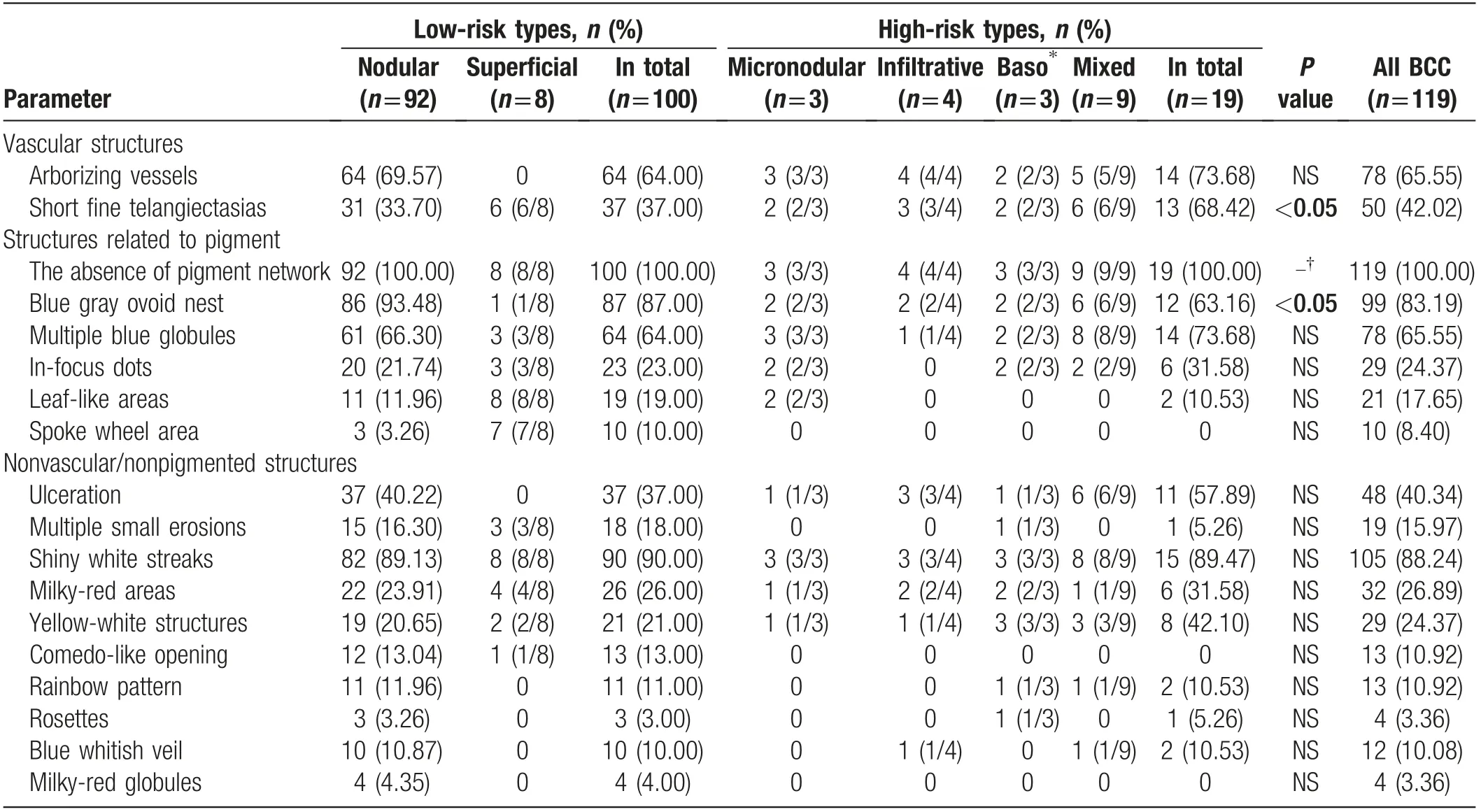
Table 1 Dermoscopic features of BCCs with low and high recurrence risks [n (%)].
Table 2 General information of patients with BCC with low and high risks of recurrence.
Histological analysis
The pathological diagnosis was obtained through surgical excision or punch biopsy. The lesions were divided into histological subtypes with a high or low risk of recurrence in accordance with the National Comprehensive Cancer Network guidelines for the treatment of cutaneous BCC in clinical practice.15Lesions with morpheaform, basosquamous (metatypical), sclerosing, mixed infiltrative, or micronodular features in any portion of the tumor were considered to have a high risk of recurrence;patients with these BCC subtypes were classified as the high-risk group.In contrast,the histological subtypes considered to have a low risk of recurrence included nodular, superficial, and other nonaggressive growth patterns, such as keratotic and infundibulocystic patterns and fibroepithelioma of Pinkus; patients with these BCC subtypes were classified as the low-risk group.
Statistical analysis
The mean age of patients in the high-risk and low-risk groups was compared using a t-test.Differences in sex and dermoscopic characteristics between the two groups were analyzed by the Chi-squared test. Differences in dermoscopic features between superficial BCC and other subtypes of BCC were analyzed by Fisher exact test.When a difference was verified, odds ratios (ORs) were calculated to understand the influence of a dermoscopic feature on the final diagnosis(identification of diagnostic predictors).Two-sided P-values of <0.05 were considered significant. All statistical analyses were performed using SPSS statistics software 21.0(IBM Corporation,Armonk,NY, USA).
Results
Clinical and demographic features of patients with BCC
Overall,119 lesions from 119 patients(63 women and 56 men) were included. The general information of patients with low-risk and high-risk types of BCCs is shown in Table 2.The sex composition and age did not significantly differ between the two groups. The most frequent anatomic location was the face (90/119, 75.63%),followed by the scalp (13/119, 10.92%), trunk (11/119,9.24%),limbs(4/119,3.36%),and neck(1/119,0.84%).Superficial BCC was more frequently located on the trunk than other types of BCC [P<0.01, OR=14.857, 95%confidence interval (CI): 3.050-72.362].
Dermoscopic features of BCC
As shown in Table 1, the most common dermoscopic characteristic of BCC was the absence of a pigment network (119/119, 100%), followed by shiny white streaks (105/119, 88.24%), blue-gray ovoid nests (99/119, 83.19%),multiple blue globules (78/119, 65.55%),and arborizing vessels (78/119, 65.55%). Yellow-white structures were present in 29/119 lesions (24.37%),including 23/119 lesions (19.33%) with milia-like cysts(MLCs),and 8/119 lesions (6.72%)with yellow lobularlike structures. Three of the “classic” BCC dermoscopic features (the six positive characteristics of Menzies’algorithm12)are listed above;the frequencies of the other three features were 17.65% (21/119 lesions) for leaf-like areas,8.40%(10/119 lesions)for a spoke-wheel area,and 40.34% (48/119 lesions) for dermoscopic ulceration.
Dermoscopic features in accordance with BCC histological type
Ninety-two lesions were diagnosed histologically as nodular BCCs (Fig. 1). The most common dermoscopic characteristic of nodular BCC was blue-gray ovoid nests(86/92 lesions,93.48%),followed by shiny white streaks(82/92, 89.13%), arborizing vessels (64/92, 69.57%),multiple blue globules (61/92, 66.30%), and ulceration(37/92,40.22%);yellow-white structures were present in 19/92 lesions(20.65%),leaf-like areas were seen in 11/92 lesions(11.96%),and a spoke-wheel area was seen in 3/92 lesions (3.26%).
Figure 1. Nodular basal cell carcinoma (BCC). (A) Dermoscopy shows arborizing vessels (red arrows), the absence of a pigment network,blue-gray oval nests(white arrows),ulceration(blue arrow),shiny white streaks(green arrow),rosettes(yellow arrows),and multiple milia-like cysts (black arrows) (x30); (B) histopathological image of a nodular BCC (hematoxylin-eosin stain, ×100).
Eight lesions were histologically diagnosed as superficial BCCs(Fig.2).Underdermoscopy,theselesionsallexhibited leaf-like areas and shiny white streaks; the other dermoscopic features included a spoke-wheel area in 7/8 lesions,short fine telangiectases in 6/8 lesions, milky-red areas in 4/8 lesions,and yellow-white structures in 2/8 lesions.No arborizing vessels or ulcerations were detected.
Three lesions were histologically diagnosed as micronodular BCCs. The dermoscopic characteristics of these lesions were arborizing vessels, multiple blue globules,and shiny white streaks.In addition,short fine telangiectases, blue-gray ovoid nests, in-focus dots, and leaf-like areas were observed in 2/3 lesions, while 1/3 lesions had yellow-white structures.
Four lesions were histologically diagnosed as infiltrative BCCs (Fig. 3). Under dermoscopy, all lesions displayed arborizing vessels, 3/4 lesions exhibited short fine telangiectases, ulceration, and shiny white streaks, and 1/4 lesions exhibited yellow-white structures.
Three lesions were histologically diagnosed as basosquamous cell carcinomas.The dermoscopic features of these lesions were shiny white streaks and yellow-white structures;in addition,2/3 lesions had arborizing vessels,short fine telangiectases, blue-gray ovoid nests, multiple blue globules, in-focus dots, and milky-red areas.
Nine lesions were histologically diagnosed as mixed BCCs(one nodular and sclerosing mixed-type lesion,and eight nodular and micronodular mixed-type lesions).The most common dermoscopic feature was multiple blue globules and shiny white streaks(8/9 lesions),followed by blue-gray ovoid nests(6/9 lesions),short fine telangiectases(6/9 lesions),ulceration(6/9 lesions),arborizing vessels(5/9 lesions), and yellow-white structures (3/9 lesions).
Dermoscopic manifestations of superficial BCC and other subtypes of BCC
As shown in Table 3, the factors significantly associated with superficial BCC were leaf-like areas(P<0.001,OR=124.037,95%CI:6.768-2273.254)andspoke-wheelareas(P<0.001, OR=252.000, 95% CI: 23.118-2746.982).In contrast, the factors significantly associated with nonsuperficial BCC were arborizing vessels(P<0.001),bluegray ovoid nests(P<0.001),and ulceration(P<0.05).
Figure 2. Superficial basal cell carcinoma(BCC).(A)Dermoscopy shows leaf-like(red arrows)and spoke-wheel areas(white arrow),and milialike cysts (black arrows) (×30); (B) histopathological image of a superficial BCC (hematoxylin-eosin stain, ×200).
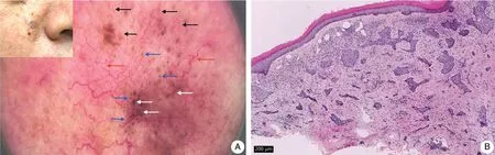
Figure 3. Infiltrative basal cell carcinoma (BCC). (A) Dermoscopy shows arborizing vessels (red arrows), short fine telangiectases (black arrows),blue-gray oval nests(white arrows),and multiple blue globules(blue arrows)(×20);(B)histopathological image of an infiltrative BCC(hematoxylin-eosin stain, ×100).
Dermoscopic manifestations of BCC subtypes with high and low recurrence risks
The dermoscopic features of BCC subtypes with different recurrence risk levels are described in Table 1.There were no significant differences between the high- and low-risk groups in the presence of most dermoscopic structures related to pigment,nonvascular/nonpigmented structures,and arborizing vessels.However,the presence of short fine telangiectases was an independent predictive factor for high-risk BCC (P<0.05, OR=3.689, 95% CI: 1.292-10.533), while the presence of blue-gray ovoid nests was an independent predictive factor for low-risk BCC(P<0.05, OR=3.904, 95% CI: 1.300-11.720). The presence of short fine telangiectases had a sensitivity of 0.684 and specificity of 0.630 for the diagnosis of highrisk BCC. The presence of blue-gray ovoid nests had a sensitivity of 0.870 and specificity of 0.368 for the diagnosis of low-risk BCC.The simultaneous presence of arborizing vessels and short fine telangiectases was a predictive factor for high-risk BCC (P=0.001, OR=6.600, 95% CI: 2.233-19.510). The sensitivity and specificity of the simultaneous presence of arborizing vessels and short fine telangiectases for predicting highrisk BCC were 0.474 and 0.880, respectively.
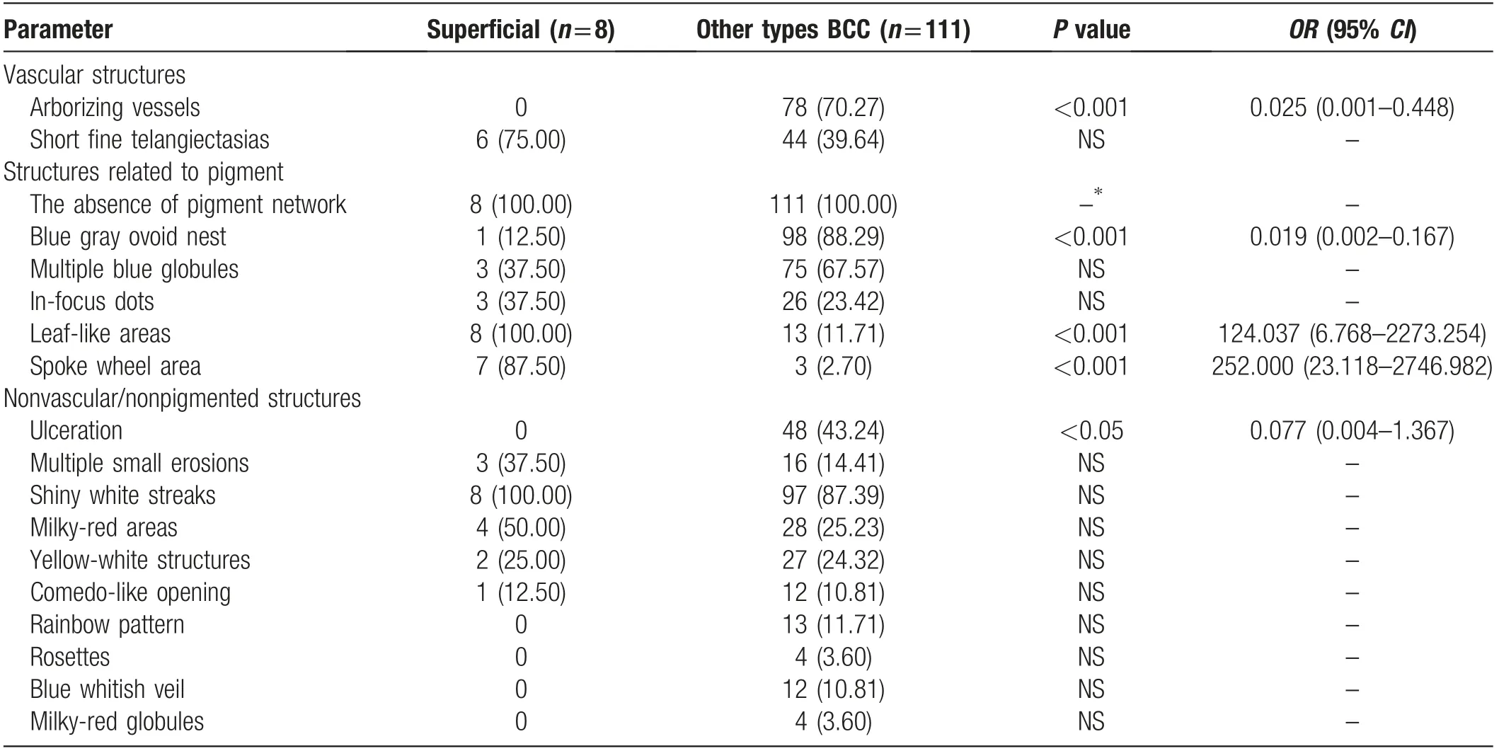
Table 3 Dermoscopic features of superficial BCCs and other histological types of BCC.
Discussion
The aim of BCC treatment is to completely remove the tumor while maximally retaining the tissue function and ensuring a good cosmetic outcome.4-5Correct judgment of the risk of pathological recurrence is important to prevent the over-removal and insufficient resection of lowrisk and high-risk BCCs,respectively.4The present results showed that there was considerable overlap in the dermoscopic features of high-risk and low-risk BCCs.However, lesions with both short fine telangiectases and arborizing vessels tended to be high-risk BCCs, with an approximately sixfold greater risk of recurrence, which may be related to the richer blood supply of high-risk BCC lesions. Additionally, blue-gray ovoid nests were an independent predictive factor for low-risk BCC. Microscopically,this structure corresponds to large well-defined tumor nests with pigment aggregates invading the dermis.10Therefore, the lower frequency of blue-gray ovoid nests in BCCs with a high risk of recurrence may be due to the smaller size and deeper location of the high-risk tumor nests compared with nodular BCCs, which accounted for the majority of low-risk BCCs.
Theyellow-whitestructuresrefertowhitetoyellow,wellcircumscribed, oval or round structures. The overall prevalence of these yellow-white structures in the present study was 24.37%,with prevalences in low-risk and highriskBCCsof21.00%and42.10%,respectively.Itshouldbe noted that small, regular, white surface scales may be mistaken for these yellow-white structures.Bellucci et al.16reported that 40/400 (10%) BCC lesions showed yellow structures,which could be divided into MLCs(7.75%)and yellow lobular-like structures (4.2%). The prevalence of these structures was higher in the present study.MLCs are common in seborrheic keratosis and congenital pigmented nevi, while yellow lobular-like structures are common in sebaceousglandhyperplasia.16-18However,ifBCC-specific dermoscopic indications are present,the possibility of BCC should not be ruled out immediately, even when yellowwhite structures are observed.16Interestingly, previous studies found an increased prevalence of hyperechoic spots under ultrasonography in histological high recurrence risk BCCs(morpheaform,basosquamous,sclerosing,etc)than histological low recurrence risk BCCs(nodular,superficial,etc).19-20These hyperechoic spots may present as yellowwhite structures underdermoscopy,and were hypothesized tobeassociatedwithseveralfeaturesofBCClesions,suchas keratosis,pigmentation,and calcification.20-21
We further analyzed the dermoscopic manifestations of different pathological types of BCC in the Chinese population. Nodular BCC was the most common pathological type, accounting for 77.31% of lesions in the present study, which is comparable to the prevalence reported in a previous study.4Consistent with a systematic review of the dermoscopic features of BCC,22the most common dermoscopic features of nodular BCC in the present study were blue-gray ovoid nests,shiny white streaks,arborizing vessels,and multiple blue globules. Micronodular BCCs had a higher prevalence of in-focus dots(66.7%,2/3),which may be related to the small size of this type of BCC.4The slightly higher prevalence of milky-red areas in invasive BCC subtypes than other BCC subtypes may be associated with the high degree of fibrosis.4,23The yellow-white structures were present in all three of the basosquamous cell carcinoma lesions in the present study, which is not consistent with the study by Akay et al.24that reported a lower prevalence(69.4%) of “white structureless areas.”
The recommended first-line options for the management of superficial BCC are nonsurgical treatments,such as imiquimod and photodynamic therapy, whereas surgical excision remains the main treatment of choice for other BCC subtypes.25-26Therefore,the present study also evaluated the difference in dermoscopic features between superficial BCC and other subtypes of BCC.Lallas et al.27reported that leaf-like areas, short fine telangiectases, multiple small erosions, and milky-red areas were potential predictors of superficial BCC. This information might be useful for clinicians to obviate the need for biopsy when planning to treat the tumor nonsurgically.27However,our results differed from these previous findings,27as only leaf-like areas and spokewheel areas were significantly associated with superficial BCCs in the present study.In addition,leaflike areas were found in 11/92 nodular and 2/3 micro-nodular BCC lesions, and a spoke-wheel area was observed in 3/92 nodular BCC lesions.Both of these features were located at the edges of the lesions. This indicates that nonsuperficial BCC may also exhibit these superficial structures when tumor nests containing pigment are located superficially at the edge of the lesion.22,28
The limitations of the present study are: (1) the small number of high-risk BCCs, and (2) the use of punch biopsy to obtain specimens for histopathological examination in a small number of cases, which may have resulted in an underestimation of some high-risk BCC lesions due to a failure to fully reflect the overall appearance of the lesion.
In summary, dermoscopy is of great importance in the preoperative evaluation of BCC in the Chinese population. Dermoscopy provides clues for the preoperative judgment of the pathological type of BCC and is helpful in differentiating superficial BCC from other BCC subtypes.Vascular structures and blue-gray ovoid nests also provide important information for the preoperative assessment of the recurrence risk of BCC lesions. The value of dermoscopy in the preoperative diagnosis of BCC is worthy of clinical promotion and further research.
Source of funding
This work was supported by grants from the Non-profit Central Research Institute Fund of Chinese Academy of Medical Sciences (No. 2019XK320024) and National Natural Science Foundation of China(Nos. 61871011 &82173449).

- 國(guó)際皮膚性病學(xué)雜志的其它文章
- Radiofrequency in Facial Rejuvenation
- Liponeurofibroma
- Successful Treatment of Severe Pityriasis Rubra Pilaris with Cyclosporine A in An Infant
- Lichen Planus Pigmentosus Inversus: Two Case Reports
- Toe Absence Related to Verrucous Carcinoma
- Dermatoscopy in the Diagnosis of Vulvar Basal Cell Carcinoma: A Case Report