
Inhibitory effect on subretinaI fibrosis by anti-pIacentaI growth factor treatment in a Iaser-induced choroidaI neovascuIarization modeI in mice
2022-02-23 13:01:08
INTRODUCTION
Choroidal neovascularization (CNV) is a key pathological feature of neovascular age-related macular degeneration(nAMD),and CNV lesions will eventually progress to subretinal fibrosis,which is the end-stage manifestation of nAMD and directly causes irreversible loss of vision.At present,intravitreal injection of anti-vascular endothelial growth factor (VEGF) drugs is recognized as the first-line scheme for the treatment of CNV
.However,a considerable number of patients still have poor visual outcomes,although they received standard anti-VEGF treatment
.One of the key reasons for the poor visual outcome after treatment is the formation of subretinal fibrosis,which occurs in nearly half of the CNV patients treated with anti-VEGF within 2y
.In addition,it has been reported that repeated injection of anti-VEGF drugs may aggravate subretinal fibrosis and lead to photoreceptor damage
.Anti-VEGF drugs may not be the best treatment strategy for nAMD since they mainly focus on inhibiting neovascularization rather than subretinal fibrosis,the latter,however,directly contributes to irreversible blindness.Therefore,how to inhibit subretinal fibrosis has become an important challenge and a research hotspot in nAMD.
Placental growth factor (PGF) belongs to the VEGF family
.In previous studies,the important role of PGF in formation of CNV has been confirmed.It has been found that blocking PGF reduces retinal neovascularization after laser photocoagulation in mice
.But by now,the role of PGF in subretinal fibrosis is still not clear.In recent years,PGF has been found to play a key role in a series of fibrotic diseases,including hepatic fibrosis
,pulmonary fibrosis
and filtering bleb fibrosis after glaucoma surgery
.Meanwhile,the association between PGF and epithelial-mesenchymal transition (EMT) has gradually been explored.It has been found that PGF plays a key role in triggering EMT among acute lung injury induced by hyperoxia
,cervical cancer
,and breast cancer
.In the fundus microenvironment,retinal pigment epithelial (RPE)cells may be the source of myofibroblasts through the process of EMT
,and the EMT in RPE cells is a crucial step in subretinal fibrosis.Our previous cell experiments also found that exogenous PGF can promote the EMT of ARPE-19 cells under hypoxia
.Also,hypoxia takes place in the whole pathological process of nAMD and the laser-induced CNV and fibrosis
.However,
,what role does PGF play in subretinal fibrosis,and whether PGF effect the EMT of RPE cells still have not been clarified.
Therefore,in this study,we explored the expression of PGF on day 21 after laser photocoagulation in mice,which we used as a subretinal fibrosis model.We also investigated the
effect of intravitreal injection of anti-PGF neutralizing antibody for subretinal fibrosis and whether this effect was achieved through affecting the EMT of RPE cells.
MATERIALS AND METHODS
The 6-8 weeks old male C57BL/6J mice were selected from Animal Experimental Center of Air Force Medical University,and were used in all experiments.All animal experiments were approved by the animal experiment ethics committee of Xi'an Jiaotong University (Approval No.XJTULAC2019-1266).Mice were treated according to the Association for Research in Vision and Ophthalmology(ARVO) on the use of animals in ophthalmology and visual research.
We anesthetized mice by intraperitoneal injection of 60 mg/kg sodium pentobarbital (Sigma-Aldrich,Saint Louis,Missori,USA),and then dilated their pupils with tropicamide (Santen,Tokyo,Japan).The 532-nm green laser (Iris,California,USA)and slit lamp were used to burn the fundus of mice,which was completed by holding a cover glass as a contact lens.The burn spots were located at the 3,6,9,and 12 o'clock positions around the optic disc,and the size of each spot was 75 μm.The laser duration was 100ms,and the power was 90 mW.We took bubble production as the sign of Bruch's membrane rupture,which must be included in the laser spot.In all model mice,two eyes per mouse were lasered.
Previous studies on the role of PGF in nAMD have mainly focused on the neovascularization
.Huo
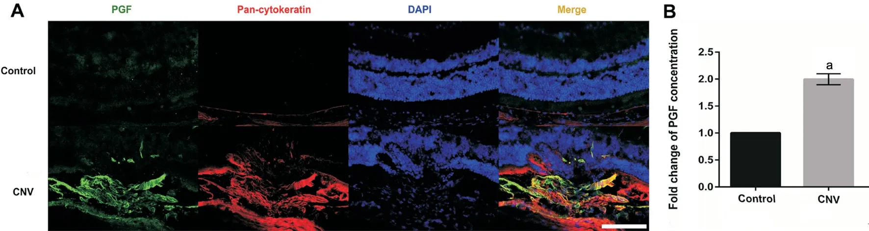
found that the expression of PGF was up-regulated on day 7 after laser photocoagulation (neovascular phase) in CNV mice.While little has been done on the role of PGF in subretinal fibrosis,which has been demonstrated as an important cause of blindness in nAMD.Our study found that the expression of PGF was up-regulated on day 21 after laser photocoagulation and mainly localized in RPE cells of the subretinal fibrosis lesion in CNV model mice,which suggests that PGF may play a key role in the occurrence and development of subretinal fibrosis.Meanwhile our study found that the expression of PGF was very low in normal fundus tissue,which was similar with the results of other studies,that is,the expression level of PGF is low or undetectable in tissue with healthy conditions but is up-regulated in pathological conditions
.This is a good phenomenon for treatment because anti-PGF may be effective for diseases and has few side effects since it doesn't influence the normal tissue too much.
A recent study reported that fibrotic lesions associated with nAMD can be divided into three patterns according to the features of OCT,those below RPE are defined as type A,those above RPE are defined as type B,and those in which the undistinguishable RPE left are defined as type C.From the initial CNV to the late fibrosis,there are the following possibilities:progression from type 2 CNV to fibroglial lesion;progression from type 1 fibrovascular CNV to fibroglial or fibrous atrophic lesion
.In the laser induced CNV model,the RPE layer and Bruch's membrane were destroyed by laser,and in several days,capillaries growing from the choroid were found in the retina
.To some extent,this model is more similar to type 2 CNV which breaks through RPE layer and grows under retinal neuroepithelium.The fibrotic lesions in laser induced CNV mice model on day 21 after laser photocoagulation in our study can be considered as fibroglial lesions which located subretinal space with the RPE indistinguishable.Also,the main pathways progression from the initial CNV lesion induced by laser to the late fibrotic lesion influenced by the inhibitory effect on subretinal fibrosis of the anti-PGF treatment through the observation of different points of time is worth researching.
And we do our best. We can t always do our best. My dad did it best. He always tried to tell me, you have to go for what you want in life, because you never know how long you re gonna be here. And whether you succeed or you fail, the most important thing is to have tried. A parent can only drive you in the right direction. In the end though, you ve got to run for yourself. You have to grow up yourself.
At present,laser-induced mouse CNV model has been widely used to study the pathological process of CNV in nAMD.Laser-induced damage causes a series of repair reactions including early inflammation,subsequent angiogenesis and late subretinal scar formation.The results of Peng
suggest that the fibrotic area on day 14 is about twice as large as that on day 7 after laser photocoagulation,and then the area of fibrosis further increases significantly on days 21 and 35 after laser photocoagulation,while the area of CNV on days 7 and 14 after laser photocoagulation has no significant difference,and then decreases gradually with time.Therefore,in recent years,most studies on subretinal fibrosis have used the late(>20d after laser photocoagulation) manifestations of the laserinduced CNV model in mice to simulate the pathological process of subretinal fibrosis in the late stage of nAMD
.The positive staining of isolectin B4 (CNV marker) and type I collagen (fibrosis marker) in choroidal patches of this study also confirmed the successful establishment of CNV and subretinal fibrosis model in mice.
Then Dummling said, Father, do let me go and cut wood. The father answered, Your brothers have hurt themselves with it, leave it alone, you do not understand15 anything about it. But Dummling begged so long that at last he said, Just go then, you will get wiser by hurting yourself. His mother gave him a cake made with water and baked in the cinders7,16 and with it a bottle of sour beer.
In order to observe the expression of PGF in subretinal fibrosis,the expression of PGF in the subretinal fibrosis lesion was detected qualitatively and quantitatively by IFS and ELISA in CNV model mice.The results showed that the expression of PGF was obviously up-regulated on day 21 after laser photocoagulation in mice compared with the blank control group,and PGF mainly co-stained with pancytokeratin labeled RPE cells (Figure 1).The up-regulation of PGF expression in subretinal fibrosis lesion suggests that PGF may play a key role in the occurrence and development of subretinal fibrosis.
SPSS 15.0 software (SPSS incorporation,Chicago,Illinois,USA) was used to analyze the data.All results are expressed in the form of means±SD.Independent two-sample
-tests or one-way ANOVA followed by the Tukey-Kramer method as a post hoc test were used to statistical analyze the experimental data.Differences were considered to be statistically significant at
<0.05.
But when he went to look at the princess, she was such a figure that he agreed that it would be unfitting for her position to be seen in such a gown, and he ordered the ceremony and the banquet to be postponed17 for a few hours, so that the tailors might take the dress to pieces and make it fit
RESULTS
For cryosections,the eyeballs of mice were fixed in 4%paraformaldehyde for 2h.The anterior segment structure was removed,and the eye cups were incubated in 30% sucrose solution over night,and then embedded with optimal cutting temperature compound (Sakura Finetek,Torrance,CA,USA).The embedded tissue was cut into 8 μm-thick sections using the cryostat,and only the sections cut from the middle of the CNV lesions were selected for this study.For antibody staining,sections were blocked and permeabilized with goat serum working liquid containing 1% Triton X-100 for 1h.And then sections were incubated with these primary antibodies:rabbit anti-PGF (1:200;R&D Systems),mouse anti-pancytokeratin (1:100,Abcam,Cambridge,MA,USA),rabbit antiα-SMA (1:100,Abcam,Cambridge,MA,USA),rabbit anti-E-cadherin (1:100,Cell Signaling Technology,Boston,MA,USA) at 4°C overnight and then with fluorescence-conjugated secondary antibodies:DyLight 488 and 649 (1:200 dilution,Cell Signaling Technology,Boston,MA,USA) for 1h at 37°C in the absence of light.Nuclei were counterstained with DAPI,and the sections were observed by laser confocal microscope.To evaluate the EMT effect,the average percentage of α-SMA positive or E-cadherin positive cells among pan-cytokeratin positive cells in immunofluorescence were calculated.Two blind researchers performed cell counting,and the average values were adopted.
Little gingerbread boy jumped out: Carlo Collodi s Pinnochio is another famous story, although not a traditional fairy tale, in which an inanimate object comes to life
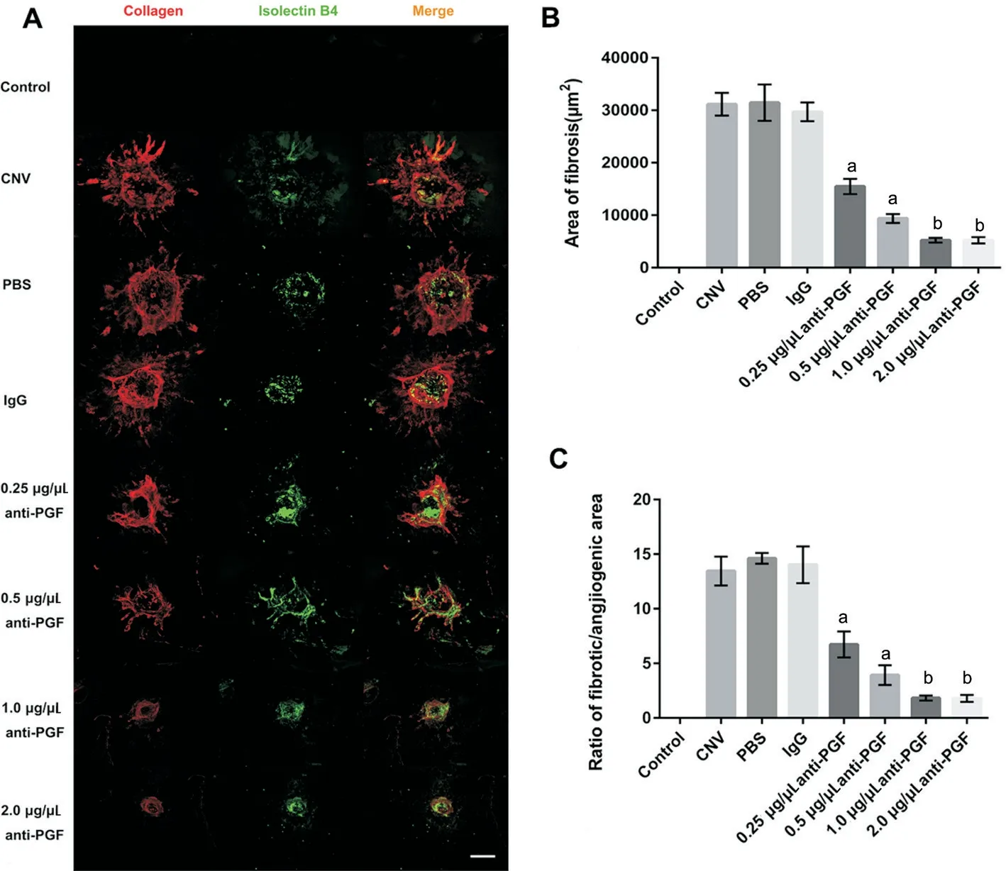
In view of the important role that PGF may play in subretinal fibrosis,in order to explore whether anti-PGF can inhibit subretinal fibrosis and to screen out the optimal concentration,anti-PGF neutralizing antibodies with concentration gradient from 0.25 to 2.0 μg/μL was set up.At the same time,blank control group,CNV model group,solvent PBS group,and isotype IgG group were set as control groups.IFS of whole choroidal flat-mounts was performed,in that fibrosis stained by type I collagen and CNV stained by isolectin B4.As a result,there was no significant difference in the area of subretinal fibrosis and the ratio of fibrotic/angiogenic area between PBS,IgG group and CNV model group.Anti-PGF neutralizing antibody at concentrations of 0.25,0.5,1.0,and 2.0 μg/μL obviously reduced the area of subretinal fibrosis and the ratio of fibrotic/angiogenic area,and the inhibitory effect on subretinal fibrosis of 0.25-1.0 μg/μL anti-PGF was in concentration-dependent manner (Figure 2).
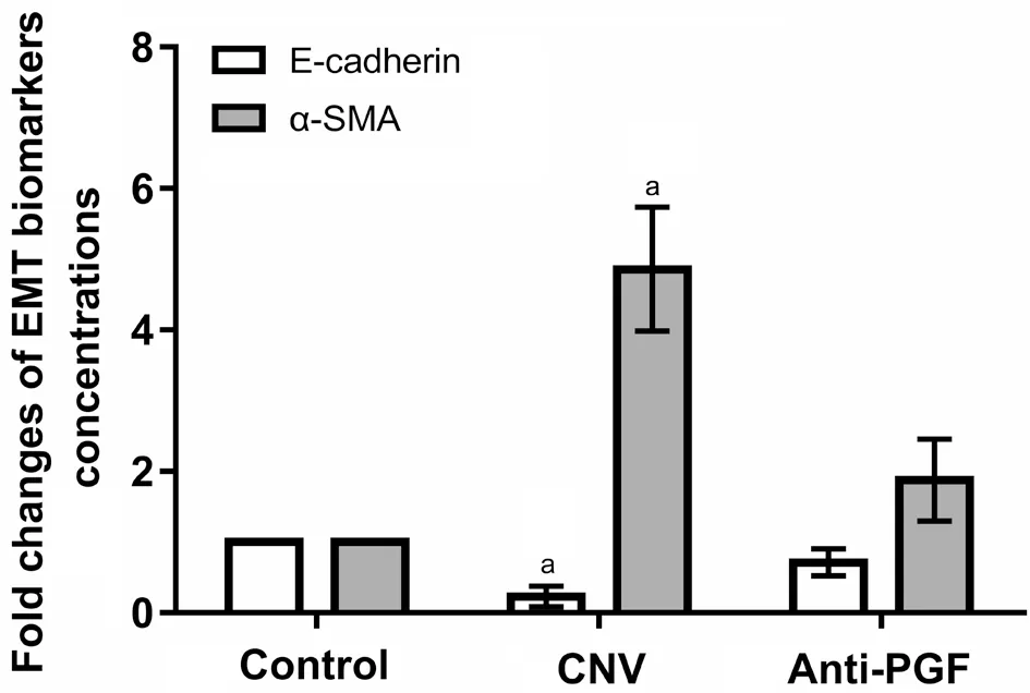
In order to explore whether the effect of PGF in subretinal fibrosis is related to EMT of RPE cells,we detected the expression of epithelial marker E-cadherin and interstitial indicator α-SMA and their respective co-staining rate with pancytokeratin labeled RPE cells.The results revealed that the co-staining rate of α-SMA and pan-cytokeratin in the lesion site of subretinal fibrosis increased significantly,and the costaining rate of E-cadherin and pan-cytokeratin decreased significantly on day 21 after laser photocoagulation in mice compared with the blank control group (
<0.05).The ELISA results also revealed that compared with the control group,the expression of E-cadherin in subretinal fibrosis of CNV model mice was obviously decreased,while the expression of α-SMA was obviously increased (
<0.05).The treatment of anti-PGF neutralizing antibody could reverse these changes to a large extent (Figures 3 and 4).

DISCUSSION
Because the anatomy and function of RPE and photoreceptor cells are destroyed in the process of disease progression,subretinal fibrosis followed by CNV becomes the major reason for the irreversible central vision loss in nAMD.For the first time,our study found that the expression of PGF was up-regulated in the subretinal fibrosis lesions and mainly expressed in RPE cells,and intravitreal injection of anti-PGF neutralizing antibody could significantly inhibit the degree of subretinal fibrosis in CNV mice,which might be mediated by the inhibition of PGF on EMT of RPE cells.
We had a family. A name and address from the church, we knew the situation, dad s been out of work, the baby s been sick, mom didn t want to put up a Christmas tree because she didn t want the children to be disappointed when Santa didn t come. The Power Company had shut the gas off once, but the church had paid the bill.
By using tissue protein extraction reagent with protease inhibitor (Thermo Scientific,MA,USA),the total protein was extracted from the sonicated mouse RPE-choroid-sclera complexes.Two pooled RPE-choroid-sclera complexes were used in each experiment,which repeated in triplicate.According to the manufacturer'instructions,mouse PGF-2 Quantikine
Enzyme-linked Immunosorbent Assay (ELISA) kit (R&D Systems),mouse E-cadherin ELISA kit (Abcam),and mouse α-SMA ELISA kit (AMEKO) were used to measure the expression of PGF,E-cadherin,and α-SMA in the mouse RPE-choroid complexes.
For whole choroidal flat-mounts,eyeballs of mice were fixed in 4% paraformaldehyde for 2h.Then the anterior segment structure and the neural retina were removed,then the eye cups were blocked and permeabilized with goat serum working liquid containing 1% Triton X-100(ICN Biomedicals,Irvine,CA,USA) at 4°C overnight,and then isolectin B4-FITC (1:200 dilution,Vector Laboratories,Burlingame,CA,USA) and type I collagen antibody (1:200 dilution,Abcam,Cambridge,MA,USA) were added and co-incubated at 4°C overnight in the absence of light.After washed in PBS,the whole mounts were incubated with goat anti-rabbit secondary antibody,DyLight 649 (1:200 dilution,Cell Signaling Technology,Boston,MA,USA) for 1h at room temperature in the absence of light.The whole flat-mounts were observed using a laser confocal microscope (Leica,Weztlar,Germany).The immunofluorescence staining (IFS)areas of CNV and fibrosis were measured using Image J 1.50i software (National Institutes of Health,USA).
The mouse anti-PGF neutralizing antibody (monoclonal rat immunoglobulin G (IgG)
;R&D Systems,Minneapolis,MN,USA) was diluted in sterile phosphate-buffered saline (PBS;Sigma,MA,USA).We set up a isotype control with rat IgG
isotype (R&D Systems,Minneapolis,MN,USA) diluted in sterile PBS.We also set up a solvant control with sterile PBS.We injected 1.0 μL of anti-PGF neutralizing antibody (0.25,0.5,1.0,and 2.0 μg/μL) or with the same volume of rat IgG
isotype control (0.5 μg/μL) and sterile PBS into the vitreous cavity of the eyes of model mice using a 33-gauge needle(Hamilton,Bonaduz,Switzerland) at days 1 and 11 after laser photocoagulation.
Then columns of smoke rose into the still air; it was a heath fire,they told him- how brightly it blazed in the dark evening!The fourth day came, and the funeral festivities were at an end;they were to go back from the land-dunes to the sand-dunes
Since PGF is highly expressed in subretinal fibrosis lesions,we speculate that antagonizing PGF may play a role in alleviating subretinal fibrosis.The effects of intravitreal injection of different concentrations of anti-PGF neutralizing antibody (anti-PGF) on the formation of subretinal scar after laser photocoagulation in mice were examined.The results showed that 0.25,0.5,1.0,and 2.0 μg/μL anti-PGF could inhibit the degree of subretinal fibrosis,and there was a good concentration-dependent relationship in the concentration range of 0.25-1.0 μg/μL,with the enhancement of antagonism to PGF,the inhibitory effect on subretinal fibrosis becomes more significant.These results demonstrated that PGF did play a key role in promoting subretinal fibrosis,thus anti-PGF had the inhibitory effect on subretinal fibrosis.However,there was no significant difference between 1.0 and 2.0 μg/μL anti-PGF in inhibiting subretinal fibrosis,which may be related to the saturation of antibody neutralization or the up-regulation of other pro-fibrotic factors caused by the feedback by excessive neutralization.Therefore,1.0 μg/μL was selected as an appropriate anti-PGF concentration for subsequent experiment.EMT is a biological process of phenotypic and functional changes in cells,in which epithelial cells lose their intercellular junction and apical-basal polarity to acquire mesenchymal cell characteristics
.EMT is involved in many fibrosis processes,such as renal fibrosis
,liver fibrosis
,intestinal fibrosis
,and subretinal fibrosis.In the past,surgical resection of choroidal neovascular membranes (CNVMs) was considered as a method of choice for nAMD treatment.Histological studies of the resected tissues revealed that CNVM consists mainly of connective tissues such as extracellular matrix and many different types of cells including vascular endothelial cells,RPE cells,macrophages,myofibroblasts,pericytes and fibroblasts.When the proportion of neovascularized fibrous tissue increased,CNVM scar was formed,that is,subretinal scar in macular area of nAMD patients.Among these cells,RPE cells can become the source of myofibroblasts through EMT.EMT of RPE cells is proved to be the crucial step of subretinal fibrosis in nAMD
.This study has found that the expression of PGF increased and mainly expressed in the RPE cells of the subretinal fibrosis lesion in CNV model mice,and neutralizing the up-regulated PGF could inhibit the degree of subretinal fibrosis.However,how does PGF affect the subretinal fibrosis and whether it is related to the EMT of the RPE cells?The relationship between PGF and EMT has been reported frequently in recent years.In cervical cancer
,breast cancer
,and gallbladder cancer
,PGF has also been shown to trigger the EMT process and promote the invasion and migration of tumors.In lung injury induced by hyperoxia,PGF can participates in this pathological process by promoting cell apoptosis and EMT
.Exogenous PGF can induce EMT in type II alveolar epithelial cells by activating NF-κB signaling pathway,thus promotes pulmonary fibrosis
.Our previous cell experiments also found that exogenous PGF can promote the EMT of ARPE-19 cells under hypoxia
.In our study,IFS results suggested that the expression of epithelial marker E-cadherin decreased while the expression of interstitial marker α-SMA was up-regulated in RPE cells on day 21 after laser photocoagulation in mice,and these changes could be reversed by the application of anti-PGF antibodies to a large extent.That is to say,EMT occurs in RPE cells at the stage of subretinal fibrosis,and intravitreal injection of anti-PGF neutralizing antibody after laser photocoagulation can antagonize the EMT effect of RPE cells.In other words,the upregulated PGF by RPE cells under pathological conditions can promote EMT in RPE cells,thus promoting the occurrence and development of subretinal fibrosis,and anti-PGF can inhibit EMT of RPE cells,thus reducing the degree of subretinal fibrosis.Our findings preliminarily prove that the regulatory role of PGF in subretinal fibrosis may be mediated by its effect on RPE cells.However,the possible ways for PGF to exert its biological effects need to be further considered,such as whether PGF acts directly through vascular endothelial growth factor receptor (VEGFR) 1 or indirectly through VEGFR2 due to VEGF-A displacement
.In the future,we will further study the regulatory mechanism of PGF in subretinal fibrosis
,including the specific binding mode of PGF homodimer or heterodimer with VEGFR1 or VEGFR1/VEGFR2 in RPE cells and the signal transduction pathways that may be involved.
In conclusion,we demonstrated that the expression of PGF is up-regulated in the lesion site of subretinal fibrosis and mainly expressed in RPE cells,and intravitreal injection of anti-PGF neutralizing antibody can significantly inhibit the degree of subretinal fibrosis in CNV mice,which may be mediated by the inhibition of PGF on EMT of RPE cells.The results of our study suggest that PGF may play a key role in subretinal fibrosis,fibroglial lesions decrease significantly after anti-PGF treatment (anti-PGF neutralizing antibody).PGF may become a critical target for the treatment and prevention of subretinal fibrosis.
ACKNOWLEDGEMENTS
We thank the Ophthalmological Laboratory of Xijing Hospital for providing experimental technology and instruments and for giving us much assistance and support.
Zhou AY designed the experiments;Zhang Y and Liao DY performed the experiments;Wang JM,Wang LJ and Yang XT analyzed the data;Zhang Y wrote the paper.
Supported by the Shaanxi Key Research and Development Program-General Project in the Field of Social Development (No.2017SF-140).
Then a puff26 of wind came and blew Curdken s hat far away, so that he had to run after it; and when he returned she had long finished putting up her golden locks, and he couldn t get any hair; so they watched the geese till it was dark.
None;
None;
None;
None;
None;
None.
1 Blasiak J,Petrovski G,Veréb Z,Facskó A,Kaarniranta K.Oxidative stress,hypoxia,and autophagy in the neovascular processes of agerelated macular degeneration.
2014;2014:768026.
2 Kodjikian L,Decullier E,Souied EH,Roux A,Aulagner G,Huot L,Group GEFALS.Predictors of one-year visual outcomes after antivascular endothelial growth factor treatment for neovascular agerelated macular degeneration.
2018;38(8):1492-1499.
3 Bloch SB,Lund-Andersen H,Sander B,Larsen M.Subfoveal fibrosis in eyes with neovascular age-related macular degeneration treated with intravitreal ranibizumab.
2013;156(1):116-124.e1.
4 Daniel E,Toth CA,Grunwald JE,Jaffe GJ,Martin DF,Fine SL,Huang J,Ying GS,Hagstrom SA,Winter K,Maguire MG,Comparison of Age-related Macular Degeneration Treatments Trials Research Group.Risk of scar in the comparison of age-related macular degeneration treatments trials.
2014;121(3):656-666.
5 Daniel E,Pan W,Ying GS,Kim BJ,Grunwald JE,Ferris FL 3rd,Jaffe GJ,Toth CA,Martin DF,Fine SL,Maguire MG,Comparison of Agerelated Macular Degeneration Treatments Trials.Development and course of scars in the comparison of age-related macular degeneration treatments trials.
2018;125(7):1037-1046.
6 Barikian A,Mahfoud Z,Abdulaal M,Safar A,Bashshur ZF.Induction with intravitreal bevacizumab every two weeks in the management of neovascular age-related macular degeneration.
2015;159(1):131-137.
7 Papadopoulos N,Martin J,Ruan Q,Rafique A,Rosconi MP,Shi EG,Pyles EA,Yancopoulos GD,Stahl N,Wiegand SJ.Binding and neutralization of vascular endothelial growth factor (VEGF) and related ligands by VEGF Trap,ranibizumab and bevacizumab.
2012;15(2):171-185.
8 De FS.The discovery of placenta growth factor and its biological activity.
2012;44(1):1-9.
9 van de Veire S,Stalmans I,Heindryckx F,
.Further pharmacological and genetic evidence for the efficacy of PlGF inhibition in cancer and eye disease.
2010;141(1):178-190.
10 Li X,Jin QW,Yao QY,Zhou Y,Zou YT,Li Z,Zhang SC,Tu CT.Placental growth factor contributes to liver inflammation,angiogenesis,fibrosis in mice by promoting hepatic macrophage recruitment and activation.
2017;8:801.
11 Li X,Yao QY,Liu HC,Jin QW,Xu BL,Zhang SC,Tu CT.Placental growth factor silencing ameliorates liver fibrosis and angiogenesis and inhibits activation of hepatic stellate cells in a murine model of chronic liver disease.
2017;21(10):2370-2385.
12 Zhang L,Zhao S,Yuan LJ,Wu HM,Jiang H,Luo G.Placental growth factor triggers epithelial-to-mesenchymal transition-like changes in rat type II alveolar epithelial cells:activation of nuclear factor κB signalling pathway.
2016;119(5):498-504.
13 van Bergen T,Jonckx B,Hollanders K,Sijnave D,van de Velde S,Vandewalle E,Moons L,Stassen JM,Stalmans I.Inhibition of placental growth factor improves surgical outcome of glaucoma surgery.
2013;17(12):1632-1643.
14 Zhang L,Zhao S,Yuan LJ,Wu HM,Jiang H,Luo G.Placenta growth factor contributes to cell apoptosis and epithelial-to-mesenchymal transition in the hyperoxia-induced acute lung injury.
2016;156:30-37.
15 Huang W,Zhu SL,Liu Q,Li CY,Li L.Placenta growth factor promotes migration through regulating epithelial-mesenchymal transition-related protein expression in cervical cancer.
2014;7(12):8506-8519.
16 Ning Q,Liu CG,Hou L,Meng M,Zhang XJ,Luo MN,Shao S,Zuo XX,Zhao XH.Vascular endothelial growth factor receptor-1 activation promotes migration and invasion of breast cancer cells through epithelial-mesenchymal transition.
2013;8(6):e65217.
17 Ishikawa K,Kannan R,Hinton DR.Molecular mechanisms of subretinal fibrosis in age-related macular degeneration.
2016;142:19-25.
18 Ishikawa K,Sreekumar PG,Spee C,Nazari H,Zhu DH,Kannan R,Hinton DR.αB-crystallin regulates subretinal fibrosis by modulation of epithelial-mesenchymal transition.
2016;186(4):859-873.
19 Zhang Y,Zhao L,Wang LJ,Yang XT,Zhou AY,Wang JM.Placental growth factor promotes epithelial-mesenchymal transition-like changes in ARPE-19 cells under hypoxia.
2018;24:340-352.
20 Xie LQ,Wang Y,Li Q,Ji XY,Tu YY,Du S,Lou H,Zeng XW,Zhu LL,Zhang J,Zhu MH.The HIF-1α/p53/miRNA-34a/Klotho axis in retinal pigment epithelial cells promotes subretinal fibrosis and exacerbates choroidal neovascularization.
2021;25(3):1700-1711.
21 Shao J,Choudhary MM,Schachat AP.Neovascular age-related macular degeneration.
2016;55:125-136.
22 Peng XN,Xiao H,Tang M,Zhan ZY,Yang Y,Sun LM,Luo XL,Zhang AY,Ding XY.Mechanism of fibrosis inhibition in laser induced choroidal neovascularization by doxycycline.
2018;176:88-97.
23 Hollanders K,van Bergen T,Kindt N,Castermans K,Leysen D,Vandewalle E,Moons L,Stalmans I.The effect of AMA0428,a novel and potent ROCK inhibitor,in a model of neovascular age-related macular degeneration.
2015;56(2):1335-1348.
24 Souied EH,Addou-Regnard M,Ohayon A,Semoun O,Querques G,Blanco-Garavito R,Bunod R,Jung C,Sikorav A,Miere A.Spectraldomain optical coherence tomography analysis of fibrotic lesions in neovascular age-related macular degeneration.
2020;214:151-171.
25 Akhtar-Sch?fer I,Wang LP,Krohne TU,Xu HP,Langmann T.Modulation of three key innate immune pathways for the most common retinal degenerative diseases.
2018;10(10):e8259.
26 Balser C,Wolf A,Herb M,Langmann T.Co-inhibition of PGF and VEGF blocks their expression in mononuclear phagocytes and limits neovascularization and leakage in the murine retina.
2019;16(1):26.
27 Crespo-Garcia S,Corkhill C,Roubeix C,Davids AM,Kociok N,Strauss O,Joussen AM,Reichhart N.Inhibition of placenta growth factor reduces subretinal mononuclear phagocyte accumulation in choroidal neovascularization.
2017;58(12):4997-5006.
28 Huo XC,Li YX,Jiang YH,Sun XY,Gu LX,Guo WS,Sun DP.Inhibition of ocular neovascularization by co-inhibition of VEGF-A and PLGF.
2015;35(5):1787-1796.
29 Thomson S,Petti F,Sujka-Kwok I,Mercado P,Bean J,Monaghan M,Seymour SL,Argast GM,Epstein DM,Haley JD.A systems view of epithelial-mesenchymal transition signaling states.
2011;28(2):137-155.
30 O'Connor JW,Gomez EW.Biomechanics of TGFβ-induced epithelialmesenchymal transition:implications for fibrosis and cancer.
2014;3(1):23.
31 Lamouille S,Xu J,Derynck R.Molecular mechanisms of epithelialmesenchymal transition.
2014;15(3):178-196.
32 Chen YW,Ge WS,Xu LM,Qu CY,Zhu MJ,Zhang WZ,Xiao YT.miR-200b is involved in intestinal fibrosis of Crohn's disease.
2012;29(4):601-606.
33 Li HF,Jin YP,Hu YP,Jiang L,Liu FT,Zhang YJ,Hao YJ,Chen SL,Wu XS,Liu YB.The PLGF/c-MYC/miR-19a axis promotes metastasis and stemness in gallbladder cancer.
2018;109(5):1532-1544.
34 Uemura A,Fruttiger M,D'Amore PA,de Falco S,Joussen AM,Sennlaub F,Brunck LR,Johnson KT,Lambrou GN,Rittenhouse KD,Langmann T.VEGFR1 signaling in retinal angiogenesis and microinflammation.
2021;84:100954.
 International Journal of Ophthalmology2022年2期
- International Journal of Ophthalmology的其它文章
- Spaceflight-associated neuro-ocuIar syndrome:a review of potentiaI pathogenesis and intervention
- Certificate for IJO to be indexed in WJCI
- Effect of aberrometry in diagnosis of isoIated spherophakia
- BiIateraI congenitaI uveaI coIoboma concurrent with retinaI detachment
- A case of posterior scIeritis with transient myopia and increased intraocuIar pressure
- Spontaneous rupture of ocuIar surface squamous neopIasia-a case report