
lntroduction of longstanding complicated sulcus intraocular lens into the intact capsular bag
2021-11-30 04:48:00FathyFawzyMorkosNaderFathyFawzyMohamedElBahrawyRaniaSeragElkitkat
Fathy Fawzy Morkos, Nader Fathy Fawzy, Mohamed El Bahrawy, Rania Serag Elkitkat,2
1Watany Research and Development Center, Watany Eye Hospital, Cairo 11799, Egypt
2Department of Ophthalmology, Faculty of Medicine, Ain Shams University, Cairo 11799, Egypt
Abstract
INTRODUCTION
Since decades, cataract has been one of the major causes of preventable loss of vision among adults worldwide[1].Following cataract extraction, an intraocular lens (IOL) should be implanted in the capsular bag for an ideal and stable visual recovery[2]. The implantation of sulcus IOLs is the second choice for many surgeons if the IOL cannot be safely implanted in the bag. Yet, this sulcus implantation is not only performed with lack of proper support for “in the bag” IOL implantation,but it can also inadvertently occur in some instances with intact capsular bag yet with a lack of surgical experience,small capsulorhexis, or narrowing of the pupil before the IOL implantation. Moreover, despite intention of “in the bag”implantation, the presence of one or both haptics in the sulcus sometimes occurs, or is discovered, postoperatively[3].
Though the sulcus implantation of IOLs is a common alternative for “in the bag” IOL implantation, there is a plethora of debate in the literature regarding the safety and stability of a sulcus implanted IOL. Though some studies support their use and report minimal complications, other studies declare significant complications that range from iris chafing, secondary uveitic glaucoma, and the uveitis-glaucomahyphema (UGH) syndrome to more serious complications like vitreous hemorrhage[4]. Furthermore, decentration of the sulcus IOLs is a fairly common complication that causes annoying symptoms like glare and monocular diplopia and can require multiple surgical trials of IOL exchange or repositioning, with both psychological and financial burdens on the patients[5-6].
The present case series study started with a case of high myopia who underwent cataract surgery 18y ago and was left aphakic, as the primary surgery was done before the era of lowpower IOLs. Our secondary surgical intervention was aiming at a surgical removal of the posterior capsule opacification (PCO)rather than using Neodymium:Yttrium-Aluminum-Garnet(Nd:YAG) laser capsulotomy, as the latter poses a higher risk of complications in aphakic eyes, including mainly vitreous protrusion and consequent retinal detachment[7]. The surgeon’s plan was to implant a sulcus IOL after complete removal of the PCO. During surgery, the idea sparkled by observing a characteristic “telltale white line” at the edge of the anterior capsule. This was supposed by the surgeon (Morkos FF) to be a landmark for the site of major adhesions that existed between the edge of the capsulorhexis and the posterior capsule that has been closed for 18y. The surgeon decided to start at this white line and try reopening the closed capsular bag, after confirming that the capsular bag was fibrotic and closed yet it was intact with no detectable capsular tears. Using combined manual and viscoelastic dissection, successful reopening of the capsular bag was achieved. Afterwards, an IOL was safely implanted inside the capsular bag. The patient was followed up and the same surgical technique was consecutively performed on other cases who experienced problems from the noxious displacements of sulcus IOLs, after confirming that they all had intact capsular bags with no detectable tears (by slit lamp examination and then confirmed intraoperatively) and hence the sulcus implantation of their IOLs was performed because of situations other than having a posterior capsular tear. This article describes the details of this proposed surgical technique and highlights the insights of its implementation for future visual rehabilitation in patients with intact yet fibrotic and closed capsular bags for years.
SUBJECTS AND METHODS
Ethical ApprovalThis is a prospective, non-randomized, noncontrolled, case series, interventional study that was performed on a recruited cohort of patients who sought medical advice at Watany Eye Hospital, Cairo, Egypt. This hospital provides primary care as well as subspecialty and tertiary referral clinics from various Egyptian governorates. The study adhered to the tenets of the Declaration of Helsinki and was approved by the Institutional Review Board of the Watany Research and Development Center (the registration number is CAT-2016-001).The participating subjects (or their guardians) were informed about the nature of the surgical technique and signed informed consents prior to the surgical procedure.
All the enrolled participants conducted the proposed surgical technique in the period from May 2016 to December 2019.The first case of this series was aphakic (as detailed above),while all the other cases had displaced sulcus IOLs that caused significant ocular symptoms of glare, visual blurring, and/or monocular diplopia. The exclusion criteria included patients with associated ocular diseases, mainly corneal pathologies,glaucoma, or posterior segment abnormalities.
For all candidates of the case series, a baseline ophthalmological examination was performed before and after the surgical intervention. This included subjective refraction [including unaided distance visual acuity (UDVA), corrected distance visual acuity (CDVA), and spherical equivalent (SE)], slit lamp examination, intraocular pressure (IOP) measurement using Goldmann applanation tonometer, and fundus examination by slit lamp fundus biomicroscopy. These candidates were also followed up (by performing the same baseline examination)for variable time intervals after the procedure.
Surgical TechniqueThe surgical technique was performed for all cases by the same surgeon (Morkos FF). Proper draping of the eye and the lashes and sterilization of the surgical field using betadine 10% was performed. This was followed by an efficient peribulbar anesthesia, except for a child who underwent the surgery under general anesthesia. Afterwards, a 1.2 mm paracentesis was performed using a clear cut blade (ALCONUSA), and a cohesive viscoelastic material (Healon GV, Johnson& Johnson, USA) was injected into the anterior chamber.
Apart from the first case of the aphakic high myope with an intact posterior capsule (described above), there was always a dislocated sulcus IOL causing significant ocular symptoms in the consecutive series of patients. These sulcus IOLs have been implanted for a range of 1mo to 7y. Following the same technique, the “telltale white line”, denoting the maximum adhesions between the edge of the capsulorhexis and the posterior capsule, was the first target for the surgeon. It was freed using combined manual and viscoelastic dissection,followed by reopening of the whole “intact posterior capsule”using the same methodology of combined dissection. As the anterior capsule started to be lifted, boluses of viscoelastic material were injected, using a fine cannula on a viscoelasticloaded syringe. The manual (mechanical) dissection of the closed capsular bag was performed using blunt dissectors, in conjunction with the viscoelastic dissection. Sharp dissection using scissors was rarely resorted to, only when there were tight adhesions, and it was cautiously performed close to the anterior capsule and guarded by injection of viscoelastic boluses. The surgeon remarkably noticed that on freeing the adhesions at the demarcated white line, the rest of the capsular bag reopened much easier than the initial reopening trials before dissecting at the white line. After the capsular bag was fully open, the displaced sulcus IOL was safely introduced into the bag in four cases while a new IOL was implanted in one case after explantation of the sulcus IOL at the beginning of surgery. Irrigation and aspiration for any newly formed lens material and/or Soemmering’s ring was then performed.Finally, the surgical technique was ended by removal of the viscoelastic substance and stromal hydration closure of the corneal incisions.
There were some special situations in some cases. In one case, the capsulorhexis was observed to be small, so it was enlarged using micro scissors to create a snip at the edge of the capsulorhexis, and then a micro forceps was used to widen the rhexis. In the same case, the IOL was found to be flipped upside down. The IOL was flipped back to its proper orientation then was safely placed in the bag after its reopening. In another case, a limited zonular dehiscence was identified preoperatively at the lower nasal quadrant, which led the surgeon to cut the preexisting sulcus IOL at its center and explant it for attaining a wider and more convenient field for intraoperative manipulations. A capsular tension ring was then placed at the defect site and a new IOL was safely implanted inside the capsular bag. In a third case, a child who had his congenital cataract removed 7y earlier presented with visual complaints from his sulcus IOL, although several surgical attempts were previously made to reposition the IOL in the sulcus. The tightly closed capsular bag was successfully reopened using a blade and vitreous scissors. A vitreous cutter was also used to extract the formed Soemmering’s ring and vitreous bands. A new IOL was then implanted into the capsular bag, which, like all the enrolled cases, was found intact yet closed for long time intervals. Composite Figure 1 shows the main steps of the proposed surgical technique.
RESULTS
This case series was performed on 6 eyes of 6 patients. The age range of the enrolled cohort was 12-68y, with mean±standard deviation of 50.8±21.19y. There were 2 males and 4 females.Table 1 shows the demographic data of the participants,their preoperative refractive condition, and the time interval between the primary surgery and the secondary intervention(which ranged from 1 up to 216mo).
Table 2 shows the refractive data of the patients before and after the surgical technique and on the follow up visits. The follow up intervals ranged between 6 and 17mo. Table 2 obviously demonstrates a significant improvement in the patients’ subjective refraction (including UDVA, CDVA, and SE) before and after surgery. It also shows a stable refractive condition for all the patients during the follow up visits.
Regarding the IOP measurements, slit lamp examination, and fundus examination by slit lamp fundus biomicroscopy, they were all unremarkable for all patients before and after the surgical procedure, apart from a tessellated fundus of high myopia in the first case with aphakia. Slit lamp examination obviously showed quiet eyes postoperatively and a centralized“in the bag” IOL in all the cases of this series.
DISCUSSION
This article presents a case series for a proposed surgical technique that enabled the reopening of intact yet closed capsular bag (with no detectable capsular tears) and the safe repositioning of the IOL from the sulcus into the bag,providing a credible stability and a secure visual quality.

Figure 1 The major surgical steps of the proposed surgical technique for introduction of longstanding complicated sulcus IOL into the intact capsular bag A: The intraoperative appearance of the sulcus displaced IOL before the beginning of the surgical procedure. B: The identification of the “telltale white line” by the surgeon (encircled in yellow), where the adhesions between the edge of the capsulorexis and the posterior capsular bag were supposed by the surgeon to be maximal, and it was a key for reopening of the capsular bag. Dissection to reopen the capsular bag began at this line.C: Mechanical dissection (using a blunt dissector) at the white line was performed. D: Viscoelastic dissection (using a fine cannula on a viscoelastic-loaded syringe) was combined with the mechanical dissection at the white line. E: Viscoelastic dissection (using a fine cannula on a viscoelastic-loaded syringe), introducing a bolus of viscoelastic material (denoted by the two yellow arrows) to separate the anterior from the posterior capsule, creating a space inside the capsular bag. F: The IOL was partially rotated into the capsular bag.The optic is now in the bag while the haptic is still in the ciliary sulcus. G:Repositioning of the trailing haptic, thus bringing the whole IOL into the fully open and intact capsular bag. H: The intraoperative appearance of the IOL while safely secured in the bag at the end of surgery.
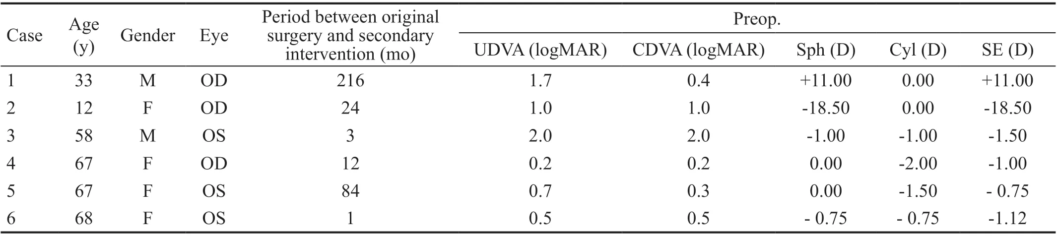
Table 1 The demographic data, period between the original primary surgery and the secondary surgical intervention, and the refractive condition of the included participants in the case series
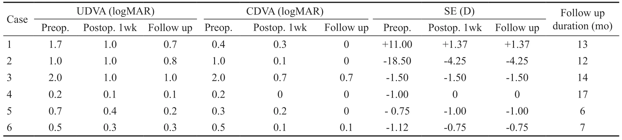
Table 2 The refractive data before and after the proposed surgical technique and on the follow up visits, together with the follow up duration of the included participants
Several trials have been conducted to safely secure an IOL inside the eye for the best visual performance and a long-term stability. The implantation of the IOL inside the capsular bag is the benchmark for attaining sustainable visual rehabilitation[2].The proposed technique in this case series provides the best scenario that a surgeon would target, which is reopening of a closed capsular bag, even with significantly long periods of closure, and an ideal implantation of the optic and haptics of the IOL in the bag. The application of this surgical technique necessitates that the patients have an intact capsular bag without capsular tears, while the IOL was placed in the ciliary sulcus for other reasons, including the lack of surgical experience, small capsulorhexis, or narrowing of the pupil before the IOL implantation. Our long follow up intervals reassured our expectations of the long-term stability of the IOL within its “presumably preferable habitat”.
To the authors’ knowledge, this surgical technique has not been previously described for reopening of an intact capsular bag that did not have an implanted IOL inside, yet the same maneuver of combined manual and viscoelastic dissection for reopening the bag was adopted decades ago during the era of frequently encountered PCO with older designs and materials of the IOLs[6]. An interesting review article by Ascasoet al[7]reported on the increasing frequency of late in-the-bag subluxation or dislocation of the IOLs after variable intervals of IOL stability, with a cumulative risk of 0.1% after 10y and 1.7% after 25y of performing the cataract surgery. The report attributed 90% of such cases to zonular insufficiency and capsular contractions, with aging, high myopia, and pseudoexfoliation syndrome being the most commonly reported risk factors. In those instances, the IOL was already implanted in the bag but had lots of surrounding fibrotic proliferations, so the reopening of the capsular bag was made easy by the existence of the IOL in the bag. In our proposed technique, the bag is wide open after being totally closed with no implantable lens inside.
The characteristic white line that the surgeon chose to be his primary target for starting the dissection was a pivotal landmark. This line represents an exaggeration of the pathological proliferation of the newly formed lens fibers and is also attributed to the phenomenon of capsular phimosis,which is an exaggerated fibrotic response that results in shrinkage of the capsulorhexis and the occurrence of adhesions between the capsulorhexis and the posterior capsular edge at the site of their contact. The exaggeration of those adhesions with an absent IOL in the capsular bag seems intuitive[8].
Regarding the presented cases in this short case series, five out of the six cases had displaced sulcus IOLs from previous surgical interventions. The causes of sulcus implantation of the IOLs varied among them. In one case, an obviously small capsulorhexis was observed and it could strongly be the cause for failure of the surgeon to place the IOL in the bag in the first surgery. Moreover, the IOL was flipped upside down in this same case, which would rather reflect the lack of surgical experience that could also lead to an inadvertent placement of the IOL in the ciliary sulcus. In another case, a limited zonular dehiscence was observed. In a third case, a child had a closed and fibrotic posterior capsule and a displaced sulcus IOL.
As we notice from the aforementioned cases of the present case series, they all had intact capsular bags that was reassured intraoperatively (even the child had a posterior rhexis that does not prevent an “in the bag” implantation of the IOL). This denotes that the IOL was inadvertently implanted in the sulcus yet having an intact capsular bag. Such cases are the ideal candidates for our described novel surgical technique. The authors strongly advise any surgeon to avoid using this technique in cases of posterior capsular rupture, as the required manipulations would rather increase the extent of the existing tear of the posterior capsule,even if small, and would result in consequent complications and failure of “in the bag” IOL implantation.
The authors believe that this novel technique provides a gateway for improving the visual performance, and hence the quality of life, for a wide cohort of patients who previously had an inadvertent implantation of a sulcus IOL yet having a good capsular bag support. This sulcus IOL has got many possible complications, and may dislodge even after several trials of repositioning, which causes both a psychological and a financial burden on those patients. Furthermore, aphakic patients who have a closed bag, especially for many years,can have a golden chance of the perfect “l(fā)ate in the bag IOL implantation”.
Worthy of mention is that the intraoperative maneuvers that were performed in the presence of a sulcus IOL require an experienced surgeon who can handle those manipulations with an existing IOL. The authors advise any surgeon who is uncertain of the proper handling of the technique with the IOL in place to remove it till completing the required reopening of the capsular bag.
The present case series is considered as a pilot study for this novel surgical technique, and the authors will retrieve more cases to perform longer case series studies in the near future.
Opening the capsular bag is not always an easy task to perform.It frequently requires patience and meticulous, even tedious,manipulations for safely reopening the posterior capsule and securing the IOL in the bag. Yet, the benefits of regaining the capsular bag for IOL implantation, especially when the patient has got a good capsular bag support from the primary surgery,undoubtedly deserve our utmost efforts.
ACKNOWLEDGEMENTS
On previous presentation of the information reported in the manuscript: Some of the cases in this article were presented as a narrated video at the American Academy of Ophthalmology,Chicago, USA, held on October 15-18, 2016. The video was also awarded as the most viewed narrated video in the conference along the following year.
Authors’ contributions:Morkos FF: proposing the surgical technique, performing the surgical procedures, clinical evaluation of the patients and their selection, and revising the manuscript and accepting it for publication; Fawzy NF: aiding the surgeon during the surgical procedures, sharing with the surgeon in the clinical evaluation of the patients and their selection, sharing in the manuscript drafting, and accepting it for publication; El Bahrawy M: data gathering, statistical analysis, sharing in the manuscript drafting, and accepting it for publication; Elkitkat RS: sharing in the data gathering and data analysis, drafting the manuscript and preparing it for publication, and manuscript submission.
Conflicts of Interest: Morkos FF,None;Fawzy NF,None;El Bahrawy M,None;Elkitkat RS,None.
 International Journal of Ophthalmology2021年11期
- International Journal of Ophthalmology的其它文章
- Toric implantable collamer lens for the management of pseudophakic anisometropia and astigmatism
- Angle-closure glaucoma with attenuated mucopolysaccharidosis type l in a Chinese family
- Novel biallelic compound heterozygous mutations in FDXR cause optic atrophy in a young female patient: a case report
- A novel temporary keratoprosthesis technique for vitreoretinal surgery
- Human umbilical cord-derived mesenchymal stem cells treatment for refractory uveitis: a case series
- Applications of dynamic visual acuity test in clinical ophthalmology