Discrepancies in the clinical and radiological profiles of COVID-19: A case-based discussion and review of literature
Hemant Kumar, Cornelius James Fernandez, Sangeetha Kolpattil, Mohamed Munavvar, Joseph M Pappachan
Hemant Kumar, College of Medical and Dental Sciences, University of Birmingham,Birmingham B15 2TH, United Kingdom
Cornelius James Fernandez, Department of Medicine & Endocrinology, Pilgrim Hospital,Boston PE21 9QS, United Kingdom
Sangeetha Kolpattil, Department of Radiology, University Hospitals of Morecambe Bay NHS Trust, Lancaster LA1 4RP, United Kingdom
Mohamed Munavvar, Department of Pulmonology & Chest Diseases, Lancashire Teaching Hospitals NHS Trust, Preston PR2 9HT, United Kingdom
J oseph M Pappachan, Department of Medicine & Endocrinology, Lancashire Teaching Hospitals NHS Trust, Preston PR2 9HT, United Kingdom
Joseph M Pappachan, Faculty of Science, Manchester Metropolitan University, Manchester M15 6BH, United Kingdom
Joseph M Pappachan, Faculty of Biology, Medicine & Health, The University of Manchester,Manchester M13 9PL, United Kingdom
Abstract The current gold standard for the diagnosis of coronavirus disease-19 (COVID-19)is a positive reverse transcriptase polymerase chain reaction (RT-PCR) test, on the background of clinical suspicion. However, RT-PCR has its limitations; this includes issues of low sensitivity, sampling errors and appropriate timing of specimen collection. As pulmonary involvement is the most common manifestation of severe COVID-19, early and appropriate lung imaging is important to aid diagnosis. However, gross discrepancies can occur between the clinical and imaging findings in patients with COVID-19, which can mislead clinicians in their decision making. Although chest X-ray (CXR) has a low sensitivity for the diagnosis of COVID-19 associated lung disease, especially in the earlier stages, a positive CXR increases the pre-test probability of COVID-19. CXR scoring systems have shown to be useful, such as the COVID-19 opacification rating score which helps to predict the need of tracheal intubation. Furthermore, artificial intelligence-based algorithms have also shown promise in differentiating COVID-19 pneumonia on CXR from other lung diseases. Although costlier than CXR,unenhanced computed tomographic (CT) chest scans have a higher sensitivity,but lesser specificity compared to RT-PCR for the diagnosis of COVID-19 pneumonia. A semi-quantitative CT scoring system has been shown to predict short-term mortality. The routine use of CT pulmonary angiography as a first-line imaging modality in patients with suspected COVID-19 is not justifiable due to the risk of contrast nephropathy. Scoring systems similar to those pioneered in CXR and CT can be used to effectively plan and manage hospital resources such as ventilators. Lung ultrasound is useful in the assessment of critically ill COVID-19 patients in the hands of an experienced operator. Moreover, it is a convenient tool to monitor disease progression, as it is cheap, non-invasive, easily accessible and easy to sterilise. Newer lung imaging modalities such as magnetic resonance imaging (MRI) for safe imaging among children, adolescents and pregnant women are rapidly evolving. Imaging modalities are also essential for evaluating the extra-pulmonary manifestations of COVID-19: these include cranial imaging with CT or MRI; cardiac imaging with ultrasonography (US), CT and MRI; and abdominal imaging with US or CT. This review critically analyses the utility of each imaging modality to empower clinicians to use them appropriately in the management of patients with COVID-19 infection.
Key Words: COVID-19; Pneumonia; Lung imaging; Chest X-ray; Computed tomography;Lung ultrasound
INTRODUCTION
The mega-pandemic caused by the severe acute respiratory syndrome coronavirus 2(SARS-CoV-2), more commonly referred to as coronavirus disease-2019 (COVID-19),continues to hit the global population even after 16 mo of its first report from China in December 2019. As of April 2021, the total death toll exceeds 2.84 million worldwide.Although COVID-19 can affect any organ system in the human body, the most clinically severe cases often have a constellation of pneumonia, acute respiratory distress syndrome (ARDS), septic shock, acute kidney injury, diarrhoea, rhabdomyolysis, and disseminated intravascular coagulation. Because of the mild nature of COVID-19 in most patients, the early diagnosis of severe illness is important for optimal treatment and appropriate utilisation of resources to prevent overstrain on the global healthcare systems. Since dyspnoea and pulmonary involvement are the most common manifestations of severe COVID-19, appropriate and early lung imaging is important not only for diagnostic evaluation, but also for prognostication. In addition, the imaging of other visceral organs may also become necessary for the diagnosis of extra-pulmonary diseases and complications related to COVID-19.
However, many of the imaging findings in COVID-19 can be nonspecific, and there can be occasional discrepancies between the imaging and clinical features seen in these patients. Moreover, on occasions, extrapulmonary disease may dominate in some patients, and pre-existing major illnesses (such as heart failure, liver diseases, chronic kidney disease and malignancies) with the acquisition of COVID-19 illness may pose additional diagnostic dilemmas. Therefore, it is important to review the sensitivity,specificity, positive and negative predictive values of each imaging technique used for the diagnosis of COVID-19 cases, and to identify the discordance that can exist between clinical and imaging features for the optimal care of patients with the disease.In this evidence-based review, we discuss these discrepancies that clinicians should be aware of, in order to manage patients with COVID-19, with the aid of three clinical case scenarios.
Case 1
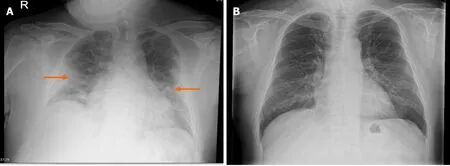
A 46-year-old male without any major past medical illness attends the emergency department (ED) with history of fever, dry cough, and intermittent dyspnoea over the past three days. His pulse oximetry showed an oxygen saturation of 92% while breathing ambient air, with an arterial blood gas analysis showing mild hypoxaemia(PaO9.3 kPa, PaCO3.82 kPa and pH 7.47). A chest X-ray (CXR) showed bilateral extensive airspace disease (Figure 1A). His full blood count showed neutrophilic leucocytosis with lymphopenia and the C-reactive protein (CRP) was elevated (232 units/L; normal < 7). The reverse transcriptase polymerase chain reaction (RT-PCR)test was positive for SARS-CoV-2 RNA confirming COVID-19 pneumonia. Management with oxygen at 2 L/minute through nasal cannula and oral dexamethasone 6 mg daily was commenced, as per the hospital protocol. This resulted in a rapid resolution of his hypoxaemia and he was discharged home on the third day of admission. A subsequent chest radiograph after 8 wk showed complete resolution of the pulmonary findings (Figure 1B).
Case 2
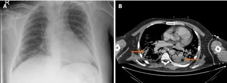
A 62-year-old lady with poorly controlled type 2 diabetes mellitus and hypertension presents at the ED with fever, loss of smell and taste, dry cough, and progressive dyspnoea in the past 2 d. Her pulse oximetry showed an oxygen saturation of 84%while breathing ambient air with an arterial gas analysis showing type 1 respiratory failure (PaO6.3 kPa, PaCO5.12 kPa and pH 7.38). A CXR was unremarkable(Figure 2A). Her full blood count showed lymphopenia without neutrophilia and CRP was 86 units/L. The RT-PCR was positive for SARS-CoV-2 RNA confirming COVID-19 infection. As the D-dimer was high (8.3 μg/mL; normal < 0.5 μg/mL) with disproportionate hypoxaemia and a normal CXR, an urgent computed tomography(CT) pulmonary angiogram (CTPA) was done which excluded pulmonary embolism(PE), but CTPA showed evidence of bilateral COVID-19 pneumonia (Figure 2B). The patient was managed with 40% oxygena Venturi mask, intravenous insulin infusion, and oral dexamethasone 6 mg daily. The patient’s hypoxaemia worsened on the next day requiring invasive ventilation and subsequently died in the intensive care unit on the 6th day with multi-organ failure.
Case 3
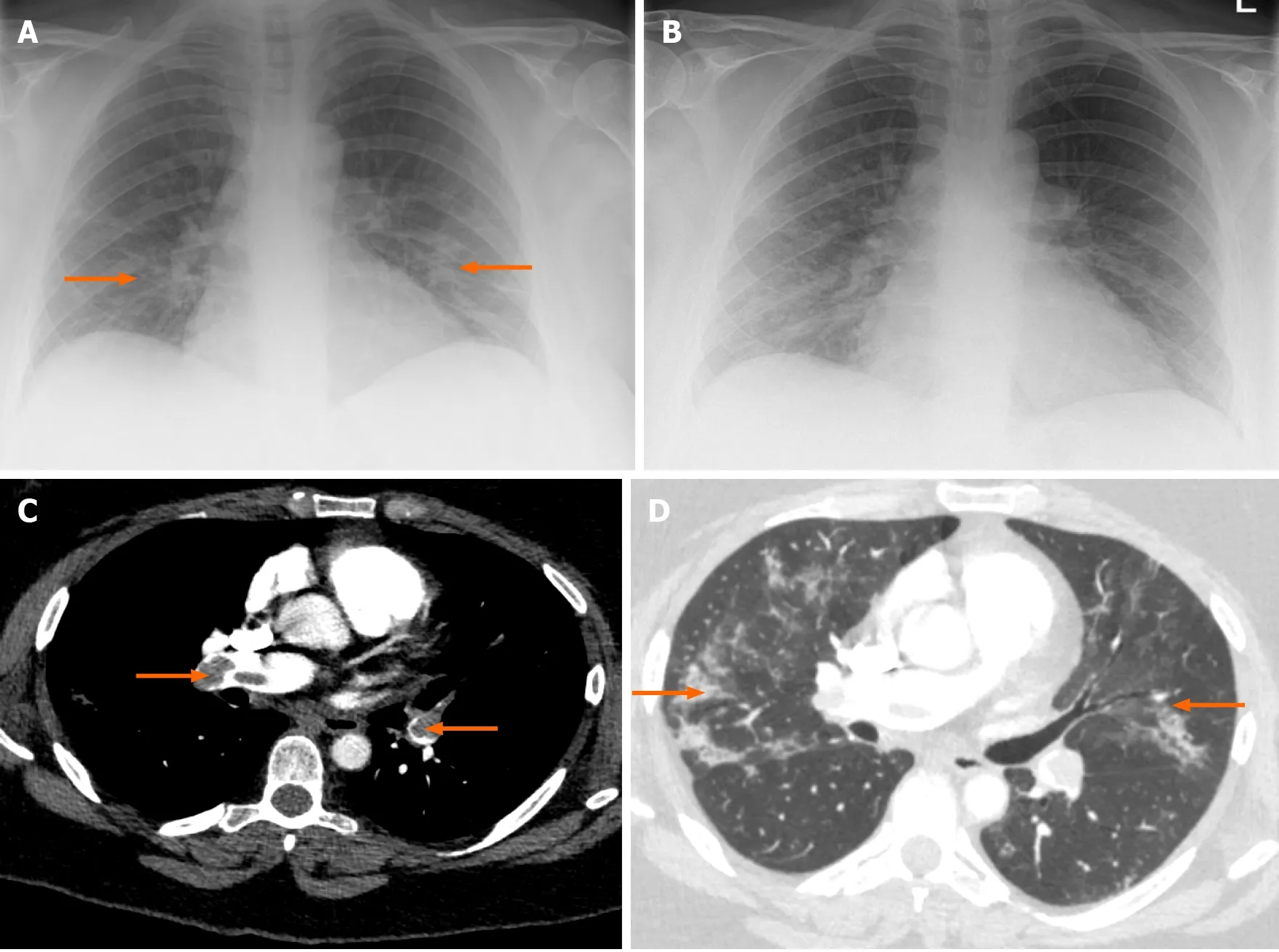
A 52-year-old lady with a history of well-controlled asthma and hypertension presented to the ED with a productive cough and breathlessness for 2 wk. Her pulse oximetry showed an oxygen saturation of 93% while on 40% oxygena Venturi mask. The arterial blood gas analysis showed type 1 respiratory failure (pH 7.43, PO7.34 kPa, and PCO5.09 kPa). There was lymphopenia (0.56 × 10/L) without neutrophilia and the CRP was 112 units/L. The RT-PCR test was positive and the CXR showed bilateral lower zone opacities (Figure 3A). She was treated with oxygen,doxycycline, and prophylactic enoxaparin and was discharged in 4 d. One week later she presented with syncope, severe breathlessness, chest tightness, tachycardia,hypoxia, and hypotension. Troponin I was raised at 205 ng/L (5-14 ng/L), along with a raised D-dimer of 6.26 μg/mL. The repeat CXR showed improving bibasilar infiltrates (Figure 3B). The CTPA showed bilateral extensive thromboembolism in the pulmonary arterial branches (Figure 3C), along with evidence of resolving COVID-19 pneumonia (Figure 3D; lung window). She underwent thrombolysis with recombinant tissue plasminogen activator, followed by therapeutic enoxaparin for an initial 4 d. She was discharged on rivaroxaban 15 mg twice daily for 21 d, followed by 20 mg once daily for 3 more mo.

Figure 1 Case 1. A: Chest radiograph (CXR) at the time of admission showing bilateral extensive airspace opacities (arrows); B: The CXR at 8 wk showing complete resolution of pulmonary opacities.

Figure 2 Case 2. A: The unremarkable chest radiograph at the time of admission; B: Computed tomographic pulmonary angiography showing evidence of bilateral coronavirus disease-19 pneumonia (arrows).
These cases illustrate some of the discrepancies between the clinical and imaging features of COVID-19. We aim to update the current evidence on the concordance and discordance between imaging and clinical profiles of the disease to empower clinicians fighting against the pandemic.
INTRODUCTION TO IMAGING STUDIES IN THE DIAGNOSIS OF COVID-19
The current gold standard for the diagnosis of COVID-19 is a positive RT-PCR test on the background of clinical suspicion. The usefulness of RT-PCR as a reference standard is limited due to its low sensitivity, sampling errors and the impact of timing of specimen collection. RT-PCR testing from a single nasopharyngeal swab in a patient with COVID-19 like symptoms has sensitivity of 87%, specificity of 97%, positive predictive value (PPV) of 98% and negative predictive value (NPV) of 80%. Because of the lower sensitivity, or when RT-PCR is unavailable, imaging tests have been used to aid in the diagnosis of COVID-19.
Another major challenge to combating the disease is its unpredictable clinical course. There is a heterogeneity in presentations, ranging from asymptomatic patients to those with critical illness, with no available prognostic biomarker to effectively triage different cohorts. Nearly 40% of transmission is attributed to asymptomatic or pre-symptomatic carriers. These asymptomatic infected individuals present unique challenges since they may not seek medical attention, thus leading to a failure of interventions that solely rely on identifying symptomatic cases. There have been some notable studies that investigate imaging in both symptomatic and asymptomatic COVID-19 positive cohorts, to assess any clinically significant findings which may guide appropriate management (discussed below).

Figure 3 Case 3. A: The chest radiograph (CXR) at the time of admission showing mild bilateral opacities (arrows); B: CXR on 2nd admission with improving bibasilar lung infiltrates; C: Computed tomographic pulmonary angiogram (CTPA) of case 3 showing evidence of extensive pulmonary embolism (arrows); D: The CTPA lung window showing resolving coronavirus disease-19 pneumonia (arrows).
Although imaging is often imperative for diagnosing, triaging and guiding management, there are some drawbacks. There may be hazards of radiation exposure,risk of COVID-19 transmission through contaminated equipment and consumption of personal protective equipment (PPE), all in the context of global resource constraints.The following section investigates the appropriate use of imaging in the assessment of different COVID-19 related pathologies, to explore the discrepancies between imaging findings and clinical presentation.
IMAGING FOR PULMONARY MANIFESTATIONS OF COVID-19
Chest X-ray
Chest radiographs are routinely used at the time of initial triage to assess for disease severity in the context of respiratory tract infections. Although systematic reviews have found that CXRs in lower respiratory tract infections do not lead to an improvement in clinical outcome, it is unclear what its role may be in COVID-19 patients.CXRs are more readily accessible than other imaging modalities and are associated with lower costs. It also allows an immediate assessment of more serious pulmonary pathologies such as pneumothorax, pulmonary oedema, pleural effusions, lung collapse and masses, although an accurate interpretation may be confounded by cardiopulmonary co-morbidities. Furthermore, the disinfection protocols for X-Ray machines are more streamlined than other bulkier systems, such as CT, or magnetic resonance imaging (MRI), which is an advantage in the era of COVID-19 disinfection protocols.
Pormohammad, conducted a systematic review to compare the laboratory and radiographic findings along with outcomes of different corona viruses [(COVID-19, SARS, and Middle East respiratory syndrome (MERS)]. 52251 COVID-19 cases,10037 SARS cases and 8139 MERS cases were included in the meta-analysis and it was seen that 85.6% of COVID-19 patients, 96% of SARS and 74% of MERS had fever at time of presentation. Cough was the presenting complaint in 63% in COVID-19 cases,54% in SARS and 61% in MERS. Severe complications such as ARDS were found in 51% of SARS, 29% of MERS and 10.6% of COVID-19 patients. With regards to chest imaging, abnormal findings were seen in a large majority of patients with all corona viruses; 84% in COVID-19, 86% in SARS and 74% in MERS. The most pertinent findings in COVID-19 patients were bilateral distribution of consolidation (76%),ground glass opacities (71%), and bilateral lung involvement (77%). Unilateral lung involvement was comparatively uncommon and was observed in 16.5% of cases.Interestingly, there were no distinguishing imaging features between different corona viruses. Between the three diseases, COVID-19 had been more contagious, thus resulted in a higher number of deaths despite its lower overall mortality rate.
As CXR can be normal in up to 63% of patients with COVID-19 related lung disease,a normal CXR does not exclude it. Typical appearances in CXR with high specificity for COVID-19 pneumonia are bilateral lower zone predominance and peripheral multifocal opacities, which could be in the form of ground glass opacities or GGO(68.5%), horizontal coarse linear opacities, or consolidation. The GGOs were shown to occur early in the disease course, and it might later progress to consolidation. The involvement is bilateral in 72.9%. However, it can be unilateral in 25% of patients with COVID-19 pneumonia. However, similar CXR findings can occur in other viral pneumonias like influenza pneumonia, organizing pneumonia or with drug reactions.Non-specific appearances that are not commonly found in COVID-19 pneumonia include unilateral perihilar opacities, diffuse involvement (without zonal preference),upper zone predominance, lymphadenopathy, cavitation, or pleural effusion with Kerley B lines.
An international panel of thoracic imaging experts released a statement during the pandemic for the use of CXR, CT and ultrasound in both suspected and confirmed COVID-19 patients. The consensus on the use of CXR is that it cannot reliably exclude COVID-19 in suspected patients, particularly in the early stages of the disease.Although CXR has a low sensitivity (69%) for the diagnosis of COVID-19 because of the non-specific imaging features, a positive CXR increases the clinical pre-test probability of COVID-19 infection. As mentioned earlier, CXRs are effective at excluding other more sinister diagnoses that may be the cause of the patient’s symptoms, which may require more immediate medical attention. CXR is not recommended for patients with mild symptoms, as it is often going to be normal in these patients, leading to a false reassurance. Thus, the timing of investigation is very important, and early investigations will often yield false-negative results. The panel also advocated a standardised reporting structure and terminology for CXRs for those with suspected or confirmed COVID-19 cases, which will be helpful in comparing findings of different studies and between different regions.
An Italian study by Schiaffino, observed that in comparison to RT-PCR for the diagnosis of COVID-19 pneumonia, CXR had a sensitivity of 89.0% (95%CI: 85.5%-91.8%), specificity of 60.6% (95%CI: 51.6%-69.2%), PPV of 87.9% (95%CI: 84.4%-90.9%),and NPV of 63.1% (95%CI: 53.9%-71.7%). A Cochrane database systematic review by Salamehobserved that, in patients with confirmed COVID-19, the pooled sensitivity of CXR is 82.1% (95%CI: 62.5%-92.7%). Another Cochrane review by Islamreported that, in patients with suspected COVID-19, the CXR has a sensitivity ranging from 56.9%-89.0% and specificity ranging from 11.1%-88.9%. However, neither group could perform a meta-analysis due to limited number of studies found.
Artificial intelligence-based algorithms have also been investigated, which showed that it can be used with higher sensitivity and specificity for the differentiation between COVID-19 pneumonia and non-COVID-19 pneumonia on CXR. In one such study by Zhang, a sensitivity of 88% (95%CI: 87%-89%) and specificity of 79% (95%CI: 77%-80%) is achieved using a high sensitivity operating threshold.Similarly, the authors reported a sensitivity of 78% (95%CI: 77%-79%) and specificity of 89% (95%CI: 88%-90%) using a high specificity operating threshold.
A study by Xiaoexplored ways to objectively assess CXRs of COVID-19 positive patients at the time of admission. The authors also assessed any correlation of CXR severity and time to intubation. Their methods involved assigning a COVID-19 opacification rating score (CORS) by dividing the lung fields on the CXR into 12 zones and counting the total number of zones showing opacity. Out of the 140 patients included in the study, 48% of patients had a CORS ≥ 6, and this was a statistically significant predictor of higher rates of intubation (OR 6.1, 95%CI: 2.1-18.1,< 0.001).Patients with CORS ≥ 6 had an intubation rate of 46%, whereas only 14% were intubated with CORS < 6. There were no significant correlations between a higher CORS and age, sex, body mass index or underlying cardiopulmonary comorbidities.Inter-rater agreement in cases of CORS ≥ 6 was noted to be moderate/substantial (κ =0.65), suggesting the scoring system was reproducible between radiographers. The findings of this study show a reliable predictor of early intubation using an objective scoring method. CORS or similar scoring criteria could have a role in reliably triaging patients with regards to planned intubation to make effective use of hospital resources such as ventilators. Although this may not correlate with clinical symptoms,it is a useful prognostic tool for risk of intubation.
Chest CT
CT scans have been shown to be a useful first-line screening method since their sensitivity has been reported as high as 98%, as opposed to the relatively low sensitivity of the RT-PCR test (can be as low as 71%). Consequently, patients are seen to exhibit positive CT findings even with negative RT-PCR tests, thus highlighting a role in addressing the RT-PCR false-negative cohort. CT chest scans have also shown to add value in prognostic information; reports show that asymptomatic cases with normal chest CTs have shorter periods from diagnosis to being COVIDnegative, than cases with positive CT findings. The limitation with this modality includes an increased exposure to radiation, higher costs, nephrotoxic contrast media,and a more time-consuming disinfection protocol.
A review into the imaging findings of COVID-19 found that CT was more sensitive than chest radiographs, and offers more unique findings which allows it to be useful even in the early disease stages. The typical findings observed include multifocal GGOs in the subpleural regions, patchy consolidation in the posterior and lower lobes which can develop into a crazy-paving pattern in later disease stages due to thickening of the interlobular or intralobular septum, and reverse CT halo sign (which are characteristic of the organizing pneumonia). Atypical findings include lung nodules,cavitation, pleural effusions, and lymphadenopathy. There have also been reports that findings of GGO are more common than consolidation in asymptomatic cases,whereas the opposite was observed in symptomatic patients. The sensitivity and specificity for the diagnosis of COVID-19 using CT scans is highest if GGO is observed simultaneously to one or more of the other aforementioned CT features (specificity:89%, sensitivity: 90%). The diagnostic odds ratio of GGO with at least one other feature is reported as 20, and is significantly higher than any sign in singularity.
Unenhanced CT chest scans may be considered as the best imaging modality to assess the extent of pulmonary involvement in patients with suspected COVID-19 pneumonia. The COVID-19 Reporting And Data System is a categorical CT assessment scheme based on unenhanced CT chest in patients with suspected COVID-19 infection, where the level of suspicion increases from very low to very high. A summary of CT observations and their interpretation, as well as classifications into various categories, is given in Table 1.
However, some suggest that CT imaging findings may not correlate with symptomatology. Nearly 54% patients showed a normal CT scan within 2 d of symptom onset, indicating that a negative CT scan should not exclude COVID-19 in patients that have relevant symptoms and exposure. In some cases, an initially negative CT scan within 2 d of symptom onset would become positive when repeated at a later stage.Another review that investigated the CT findings in asymptomatic COVID-19 positive cases showed that among 63% of patients who had positive CT findings, 58%remained asymptomatic, however the remainder developed the symptoms. Out of those who developed symptoms, 90% had previously shown positive CT scans. This important study proves the need for close clinical monitoring of asymptomatic cases with radiographic findings since a significant percentage will become symptomatic.
Although CT scans are more effective in identifying COVID-19 related changes than CXR in the early disease stages, it still has limited sensitivity and negative predicative value for ruling out COVID-19 infection. A Cochrane database systematic review by Salamehobserved that, in patients with confirmed COVID-19, the CT chest has a pooled sensitivity of 93.1% (95%CI: 90.2-95.0), though the studies had considerable heterogeneity. However, in suspected cases where CT scans were used as a first-line diagnostic test, the sensitivity was 86.2% (95%CI: 71.9-93.8), and the specificity was only 18.1% (95%CI: 3.71-55.8) with a high degree of heterogeneity. A subsequent Cochrane review by the same group observed similar findings with a pooled sensitivity of 89.9% (95%CI: 85.7-92.9), and a pooled specificity of 61.1% (95%CI: 42.3-77.1), in patients with suspected COVID-19. These systematic reviews indicate that,in suspected patients, CT chest may not be able to differentiate COVID-19 from other respiratory illnesses. Therefore, CT chest should not be used as a stand-alone tool foran early assessment of COVID-19 status and, in an ideal setting, may be best used in combination with RT-PCR. This would accommodate for the low sensitivity of RTPCR, thus giving a more holistic approach for COVID-19 diagnoses. Similarly, CT chest scans can also be used in those patients with negative RT-PCR, but with typical symptoms of COVID-19, to address the false-negative cohort.

Table 1 Coronavirus disease-19 Reporting and Data System category of computed tomography reporting and the level of suspicion for coronavirus disease-19[41]
The reduced specificity of the CT scans could partly be due to the lower sensitivity of the reference standard (RT-PCR). A systematic review by Kovács, observed that CT chest has a high sensitivity (67%-100%), and low specificity (25%-80%),whereas the RT-PCR has only a modest sensitivity (53%-88%). They applied a reverse calculation approach, after considering CT chest as a hypothetical gold standard. The reverse calculation approach showed that CT could have a higher specificity (83%-100%), once the modest sensitivity of RT-PCR is considered. Similarly, an Italian study by Falaschi, observed that, when compared to RT-PCR, CT chest has a sensitivity, specificity, PPV, NPV, and accuracy of 90.7%, 78.8%, 86.4%, 85.1% and 85.9%,respectively. A recent French study by Herpe, observed that CT chest has a sensitivity of 90% (95%CI: 89%-91%), specificity of 91% (95%CI: 91%-92%), PPV of 89%(95%CI: 87%-90%), and NPV of 92% (95%CI: 91%-93%), when compared to RT-PCR.
Furthermore, CT scans can be utilised to closely monitor disease progression since imaging features have been observed to follow a predictable time course. Following symptom onset, the severity of CT findings progresses rapidly to peak at 6-11 d, from which it shows slow resolution, and the disease course can be classified into four stages. In the early stage (0-4 d), GGOs are seen in the subpleural lower lobes. This is followed by a progressive stage (5-8 d) where multiple, diffuse GGOs are seen in bilateral lobes with crazy-paving and /or consolidation. In the next peak stage (9-13 d), there are extensive GGOs with dense consolidation and crazy paving pattern with or without parenchymal bands. Lastly, the absorption stage (over 14 d) shows gradual resolution with an absence of crazy paving. These stages have been echoed by other studies as well.
In addition to the four similar temporal stages of CT findings, Kongnoted the presence of fibrosis-like strips in the lower lobes and their median time of appearance was 4.5 d after disease onset. Among 88% of the cases, these strips disappeared within 9-20 d. This study also noted a correlation between symptomatology and CT findings. Nearly 14/22 patients had resolution of fever during the progressive stage, and all patients that had cough showed resolution during the last‘dissipation stage’ (akin to the above absorption stage). Since this was a small study,correlations between clinical and imaging findings cannot be conclusively stated.
A semi-quantitative scoring system was developed to correlate the extent of pulmonary involvement with the clinical stage of the disease and laboratory findings to assess the role of CT in predicting short-term outcomes including mortality. The scoring system involved considering the extent of anatomical involvement to each of the 5 lung lobes, with each lobe involvement being visually scored from 0 to 5, as given in Table 2. This was then combined to get a sum for a total CT score that ranged from 0 (no involvement) to 25 (maximum involvement). Francone, observed that the CT score to be significantly higher in critical and severe cases compared to other groups. Moreover, the CT score was significantly correlated with CRP and D-dimer levels. A total CT score of over 18 was associated with an increased mortality risk andwas predictive of death in univariate analysis and multivariate analysis [HR 8.33(95%CI: 3.19-21.73;< 0.0001), and 3.74 (95%CI: 1.10-12.77;= 0.0348) respectively].This system, although reliant on subjective CT reporting, shows how CT can be used for prognostication. Inter-rater agreement should be measured in future studies to understand the reliability between different reporters and institutions.

Table 2 Extent of lung involvement and the corresponding computed tomography scores[47,49]
Pan, studied the extent of lung involvement using the total CT score in various stages of the diseases and they observed that the total CT score peaked at the 10day of illness. In their study the total CT score in stage 1 (early stage) was 2 ± 2, in stage 2 (progressive stage) it was 6 ± 4, in stage 3 (peak stage) it was 7 ± 4, and in stage 4 (absorption stage) it was 6 ± 4. Moreover, the lung involvement increased, and consolidation was observed up to 2 wk after the onset of the disease. Thereafter,consolidation was absorbed, leaving extensive GGO and sub-pleural parenchymal bands.
Patients with severe COVID-19 infections requiring hospitalization (even those who are not critically ill) are at high risk of venous thromboembolism (VTE), and a significant proportion would develop VTEs even while on standard prophylactic anticoagulation therapy. The incidence of VTE in patients admitted to the intensive care unit, even after receiving standard prophylactic anticoagulation therapy with low molecular weight heparin (LMWH), is nearly 25%. A recent study by Jalaber,observed that the prevalence of acute pulmonary embolism at initial presentation in unselected (severe and non-severe) COVID-19 patients is less than 6%. They observed that most patients who developed acute pulmonary embolism had D-dimer levels above 5000 ng/mL. Moreover, the median interval between symptoms onset and pulmonary embolism was 7 d. Thus, acute PE occurred in the second stage of COVID-19 illness corresponding to the cytokine storm. Current explanations for the pathogenesis of COVID-19-associated hypercoagulability include hypoxia and systemic inflammation secondary to COVID-19 that may lead to high levels of inflammatory cytokines and the consequent activation of the coagulation pathway.However, the exact mechanisms causing thromboembolic episodes remain elusive.Though disseminated intravascular coagulation was suggested as one of the pathogenic mechanisms, a recent study by Martín-Rojas, has not shown an evidence for consumption coagulopathy in these patients.
A recent National Institute for Health and Clinical Excellence guideline recommends initiating standard prophylactic anticoagulation with LMWH, fondaparinux, or unfractionated heparin, and to continue them for the duration of hospitalisation or 7 d,whichever is longer. Patients requiring advanced respiratory support in the form of invasive mechanical ventilation, bi-level positive airway pressure, continuous positive airway pressure or extracorporeal membrane oxy-genation can be considered for doubling the standard prophylactic dose of LMWH, which is also known as intermediate-prophylactic dose (off-label indication).
Routine use of CTPA as the first-line imaging modality in patients with suspected COVID-19 pneumonia is not justifiable due to the deleterious effects of contrast administration in patients at high risk of developing acute kidney injury (particularly those that are elderly and those with diabetes). Artificial intelligence-based algorithms have been developed to evaluate diagnostic test accuracy of CT chest in diagnosing COVID-19 lung disease. A recent study using multinational database found that artificial intelligence-based algorithms exhibited 90.8% accuracy, 84% sensitivity and 93% specificity in the detection of COVID-19 pneumonia in the CT chest, indicating that these algorithms can readily identify COVID-19 pneumonia in CT chest imaging,and can distinguish non-COVID-19 related pneumonias with high specificity.
Lung ultrasound
The use of lung ultrasound (LUS) is gaining interest in the context of COVID-19.However, its usefulness in diagnosis is still uncertain. The modality has unique advantages since it is quick and cheap to use, non-invasive, easily accessible, easily sterilised, and it does not involve radiation. It also allows an immediate exclusion of other causes of symptomatology, including myocardial injury and pneumothoraces.This bedside test also negates having to move infected patients from their isolated hospital areas (, into CT scanners), and offers a rapid assessment of lung infection,and also offers an alternative investigation to those who cannot accept CT scans (, in pregnant patients). However, it is reliant on an operator with sufficient expertise, and it is difficult to gain useful information about deeper structures. As with CT scans, the sensitivity and specificity are thought to increase with severity of infection. In a Cochrane Database systematic review, it was observed that in suspected COVID-19 patients, the LUS has a sensitivity of 96.8% and a specificity of 62.3%.
It is reported that the evaluation of respiratory failure in critically ill patients is better done with LUS than CT or CXR, especially in the context of ARDS and pneumonia. LUS can also be used to define severity and progression of COVID-19 associated lung changes, with different stages of the disease being characterised by typical findings. On US scans, normal reverberation artifacts of pleural lines show as motionless and regularly spaced lines perpendicular to the pleura - these are A-lines.From the pleural lines, vertical linear artifacts which arise from thickened interlobular septa and other subpleural structures represent B-lines. The number of B-lines increases as the lung density increases and with reduced air content, which indicates pulmonary interstitial pathology. Mild-moderate disease will show scattered B lines,thickened pleural lines, skip lesions (alternating areas of B lines with A lines), along with small consolidations (around 1 cm). In severe disease, B lines become confluent,and consolidation increases. Furthermore, in critically ill COVID-19 patients, extensive coalescent and disappearing B lines affecting upper and anterior lungs, with corresponding consolidation are observed. Apart from the physiological A lines,and pathological B lines, other artifacts such as C, E and Z lines, have been described. Moreover, as consolidation increases, the echogenicity of the lung changes to resemble that of the liver (‘hepatic pattern’), while also demonstrating changes to the bronchi morphology. The ‘shred sign’ denotes the shredded appearance at the interface between consolidation and normal lung; this is thought to be the most specific sign of pneumonia in LUS.
According to most studies, these findings are non-specific and there are no pathognomonic LUS signs of COVID-19 that can be used to establish a definitive diagnosis. In some studies, however, point of care ultrasound (POCUS) of the lungs done in the emergency department exhibited a strong correlation with CT scan findings in COVID-19 patients. A recently published study by Haak, showed that the POCUS has an overall sensitivity of 89% (95%CI: 70%-97%), specificity of 59%(95%CI: 46%-71%), PPV of 47% (95%CI: 33%-61%), and NPV of 93% (95%CI: 79%-98%).In patients without past medical history of cardiopulmonary disease, POCUS has a sensitivity of 100% (95%CI: 70%-100%), specificity of 76% (95%CI: 54%-90%), PPV of 67% (95%CI: 41%-86%), and NPV of 100% (95%CI: 79%-100%).
In addition, Millingtonestablished four probabilities of disease manifestations on LUS. High probability LUS patterns include bilateral and multifocal clusters of Blines, alternating with normal lung, with some peripheral consolidation. Intermediate LUS patterns involve unilateral clusters of B lines with or without peripheral consolidations. Alternate LUS patterns are observed with large consolidation or effusions and symmetrical B-lines. This may suggest alternative diagnosis such as cardiogenic pulmonary oedema. Low probability LUS patterns is characterised by generalised bilateral A-line pattern and may also indicate an alternative diagnosis.LUS is observed to have positive findings in early stages of pneumonia, and before disease symptomatology. Extrapolating this further, US may have a role in early diagnosis and monitoring of COVID-19 positive patients, especially in the critical care setting and in pregnant women, where there is a high threshold for the use of other modalities. Table 3 compares the sensitivity, specificity, PPV and NPV of various lung investigations that were used in the diagnosis of COVID-19 infection, where RT-PCR is considered as the gold-standard and the other modalities are compared to it.
MRI
Thoracic MRI can be used as a radiation-free tool for the evaluation of lung parenchyma in high-risk patients with COVID-19 infection including pregnant women and children, in whom ionizing radiation should be avoided. Movement artfacts and thelow proton content of normal lungs make its evaluation by MRI difficult. However,the consolidation and ground-glass opacities occurring in patients with COVID-19 pneumonia cause an increase in proton content and signal intensity of the lungs.Moreover, advanced technologies such as T2-weighted spin-echo PROPELLER MRI sequences provide rapid acquisition of images, with better quality and with lesser artefacts, in as quick as 3 min.

Table 3 Efficacy of reverse transcriptase polymerase chain reaction, chest X-ray, computed tomography chest, and lung ultrasound in the diagnosis is of coronavirus disease-19 [16,24,43,44,69]
A study that compared the findings on CT chest and MRI chest in patients with COVID-19 infection revealed that 90% of patients had ground-glass opacities in CT and MRI. Consolidation was observed in nearly 45% of patients in both CT and MRI.Moreover, nodules were detected in 37.5% by CT and in 34.4% by MRI. In summary,when compared to CT chest, in the detection of lung nodules, MRI chest has a sensitivity of 91.7%, specificity of 100%, positive predictive value of 100%, and negative predictive value of 95.2%. Another study, in which ultrashort echo time MRI was compared with conventional CT imaging in patients with COVID-19 infection,showed that there is substantial agreement for detecting the typical lesions of COVID-19 pneumonia, including pure GGOs, pure consolidation, and GGOs with consolidation.
Myocardial injury characterized by troponin elevation is seen in nearly 17% of patients with COVID-19 infection. Many of these patients might develop new onset left ventricular systolic dysfunction of uncertain aetiology, as observed on fast bedside echocardiography. The cardiovascular magnetic resonance (CMR) done in these patients might also show interstitial myocardial oedema, contractile dysfunction,cardiomyocyte necrosis, or myocardial fibrosis. The pulmonary magnetic resonance angiography (MRA) might facilitate direct visualisation of filling defects in the pulmonary arteries and/or as parenchymal perfusion defects. The CMR protocol for the cardiovascular structures can be combined with the MRI protocol for the pulmonary arterial tree and lung parenchyma; this combined cardiothoracic-MRI protocol might save precious time in these critically unwell patients. Cranial MRI is a useful investigation in the evaluation of anosmia.
Positron emission tomography
The positron emission tomography (PET) scan is not considered as one of the primary diagnostic modalities in patients with COVID-19 infection, even though it possesses high sensitivity to detect subclinical infection. The disadvantages of PET include low specificity, high cost, prolonged exposure time, and time-consuming decontamination process. However, prompt detection of COVID-19 infection and early initiation of supportive treatment in patients undergoing PET for other indications including malignancies and other immunocompromised states could improve survival.
IMAGING FOR THE EXTRA-PULMONARY MANIFESTATIONS
Neuroimaging
COVID-19 infection also has impacts on the nervous system. Patients often present with anosmia, ageusia, headache, stroke, impairment of consciousness, seizures, and encephalopathy. These nonspecific manifestations warrant cranial imaging with CT or MRI to exclude severe acute manifestations including intracranial haemorrhage,cerebral venous sinus thrombosis, stroke, and encephalomyelitis. A systematic review of five observational studies found that 24% of COVID-19 positive patients presented with neurological features, while not suffering from any respiratory symptoms. The pooled incidence of acute stroke was estimated to be 1.2% with a high mortality rate of 38% in these patients. The mean age of patients considered was 63.4 years and it was thought to be difficult to establish a causal relationship between COVID-19 and cerebrovascular stroke due to co-existent cardiovascular comorbidities.In addition, clinicians need to be aware of immune mediated complications such as Guillain-Barre syndrome and acute disseminated encephalomyelitis. Mood disorders and psychosis are also some of the documented neuropsychiatric disorders associated with COVID-19.
Although the neurological symptoms may be quite dramatic, the imaging may not show any positive findings. If present, positive findings on cranial imaging include evidence of cerebrovascular diseases with haemorrhagic or non-haemorrhagic infarction, cerebral venous thrombosis, thrombotic microangiopathy, infectious and inflammatory diseases, acute disseminated encephalomyelitis and other demyelinating diseases including white matter demyelinating diseases. Patients with severe COVID-19 infection and subsequent intensive care unit admission may develop neurological sequalae; this is thought to be due to brain ischaemia, however, one must rule out the possibility of chronic neuro-inflammation and demyelination during follow up appointments of recovered patients. Hence, neuro imaging should be organised to exclude demyelinating diseases including multiple sclerosis.
Cardiovascular imaging
Cardiovascular complications were demonstrated in various studies in patients affected by COVID-19. Appropriate multi-modality imaging should be considered depending on the clinical manifestations of the disease, which are all part of a nonspecific spectrum of symptoms. Considering the easy availability and feasibility of bedside US scans, any patients with suspected cardiovascular symptoms should be directed to a preliminary echocardiography to assess for ischaemic or non-ischaemic myocardial injury and pericardial pathologies. The clinical severity of cardiovascular symptoms helps to determine the appropriate imaging and interventions, with consideration to the laboratory investigation results. Mild symptoms warrant bedside US scans which have the advantage of being radiation free, portable, repeatable, quick,and inexpensive and can be performed in isolation with minimal risk of spread of infection.
In an acute clinical set up, the role of cardiac CT and MRI are reserved for patients with more serious or inconclusive symptomatology and the evidence for their use is still mounting. Although chest CT scan provides detailed information on the mediastinum and the lungs, early changes of myocardial oedema is best appreciated on cardiac MRI. The use of cardiac imaging by CT and MRI are limited by their availability, time, cost, and patient factors. It is also recognised that comorbid cardiac pathologies complicate the clinical picture of COVID-19 positive patients. In addition to worsening the pre-existing cardiovascular disease, COVID-19 infection can cause primary infective/inflammatory changes in myocardium and pericardium with possible myocarditis, myopericarditis, myocardial ischaemia and pulmonary emboli.
Abdominal imaging
Approximately 16% of COVID-19 patients present with gastrointestinal symptoms with or without respiratory symptoms. In one study by Behzad, 0.5%-19% of patients showed some form of renal dysfunction, and it is observed that renal dysfunction in COVID-19 can be caused by a direct endothelial invasion of the glomerular capillaries (endotheliitis). The knowledge of this is particularly important when planning a contrast enhanced CT scan or MRI due to the risk of inducing contrast nephropathy. Several COVID-19 patients demonstrated abnormal liver function but there are no specific diagnostic features. Imaging by US scan and CT showed hepatic steatosis, which is also a nonspecific finding, which could be attributed to pre-existing changes or even due to drug induced pathologies. In more severe COVID-19 infection, multi organ disease can also manifest as septic shock,refractory metabolic acidosis, and coagulation dysfunction.
It was also observed that COVID-19 infected patients shed detectable traces of virus in faeces. It is found that the stool samples remain positive for viral RNA after respiratory samples became negative. This implies that the health care workers dealing with suspected or confirmed COVID-19 patients requiring endoscopy and collecting faecal samples during patient recovery should be done with proper use of PPEs. On CT imaging of the abdomen in COVID-19 positive patients, the nonspecific findings include small and large bowel wall thickening, fluid filled colon, pneumatosis intestinalis, pneumoperitoneum, intussusceptions, and ascites. A small study in COVID-19 infected pregnant ladies showed that there are no specifically different aspects of the disease and the findings are just as nonspecific as in the general population.
DISCUSSION
Why can there be clinical and imaging discrepancies among COVID-19 patients?
CT chest images of some COVID-19 patients showed apparent lesions in the lungs, yet the RT-PCR repeatedly showed negative results until eventually turning positive.This discrepancy is due to the increased sensitivity of CT chest compared to RTPCR. Among imaging studies themselves, there exist discrepancies in the diagnostic yield, especially when imaging is done in the early stages and in patients with mild COVID-19 infections. Moreover, interstitial pneumonia is one of the most common features of COVID-19. The CXR is less sensitive in detecting interstitial pneumonia,in comparison to high resolution CT chest. There is a high thromboembolic risk associated with COVID-19. Many of these patients may present simply with dyspnoea and type 1 respiratory failure where a normal CXR or non-contrast CT chest often would not exclude pulmonary thromboembolism. Prompt evaluation with CTPA or MRA should be considered when such clinic-imaging discrepancy is present.
Certain patients with extensive pneumonic changes in CXR will come off oxygen and could be discharged soon after admission, whereas some other patients without extensive pneumonic changes succumb to the disease. This is mostly related to the varied propensity to develop the cytokine storm syndrome, which is responsible for acute respiratory distress syndrome, multiorgan failure and death.
Discrepancies among children and young adults
The clinical and imaging features of COVID-19 can be different from that of middleand old-age adults. This is often related to milder course of the clinical illness in most of these patients. Consolidations with surrounding halo signs were peculiar to paediatric COVID-19 pulmonary disease, probably resulting from underlying coinfectionswhereas the imaging features were different among older adults as described earlier. Paediatric cases less commonly show a peculiar presentation of COVID-19 known as multisystem inflammatory syndrome in children (MIS-C). The chest radiographic features of MIS-C include pleural effusions and opacifications of lower lung zones.
Young adults and adolescents are usually affected by less severe attack of SARSCoV-2 virus compared to middle aged and older adults. Therefore, the clinicoradiological features of such patients are expected to be different from those among older adults. This discrepancy could be explained by more profound virus-related immune dysfunction in the latter age-groups and the associated co-morbidities.There seems even a gender preponderance in the severity of COVID-19, with males affected more than the females, necessitating longer periods of hospitalisation,intensive care unit admissions, and mortality.
CONCLUSION
Although the advent of RT-PCR testing for COVID-19 has undoubtedly been crucial in combating the disease, its diagnostic utility remains limited due to its relatively low sensitivity. For this reason, imaging tests are now gaining focus to aid the diagnosis in clinical scenarios where the RT-PCR result is inconclusive. Since most symptoms relate to the respiratory system, the use of chest imaging plays a crucial role in diagnosis.CXRs are a quick and useful tool for an immediate assessment of thoracic pathology.However, it must be noted that its sensitivity for diagnostic purposes is far from perfect, owing to the lack of specificity in findings seen in COVID-19. It must be noted,however, that chest radiographs with COVID-19 opacification rating score is a significant predictor of the need for intubation, while exhibiting reproducibility between reviewers.
CT scans, on the other hand, show good sensitivity compared to RT-PCR, however its specificity is less. Combined observation of GGO and one other sign of pneumonia(septal thickening, consolidation, or pleural effusions) is the most specific and sensitive sign for COVID-19. CT has been shown to be useful for disease monitoring as it is can be used to map the disease in its temporal stages. The advantage of CT chest is that it gives both diagnostic and prognostic information. However, the limitations with CT chest (either unenhanced or CTPA) includes the cost, increased radiation exposure,and the difficult as well as time-consuming disinfection protocols. LUS has been proven to be superior to CT and CXR in the assessment of critically ill patients,especially with ARDS and pneumonia and can be used to monitor disease progression,severity, and resolution. Again, the findings are non-specific and there are no pathognomonic findings to indicate a COVID-19 diagnosis. MRI of lungs has recently emerged as a highly sensitive and specific imaging tool for diagnosis of COVID-19 lung disease especially when CT scans pose higher radiation risk among children,adolescents and pregnant women.
With regards to extra-pulmonary manifestations of COVID-19, imaging plays a key role in identifying and monitoring these impacts. Neurological sequelae are common and range from ischaemic injury to demyelinating conditions. Thus, neurological imaging is paramount in those with symptoms, and it is equally important to use them during follow up. Cardiac imaging can be in the form of echo, or cardiac CT/MRI, and the symptom severity and clinical suspicion of an underlying cardiac diagnosis will guide clinicians to use a specific modality. Abdominal symptoms are often present in COVID-19 patients and require monitoring. A significant proportion of patients may also suffer from renal impairment. Thus, imaging and laboratory tests are needed to monitor this, especially since contrast enhanced imaging is often required for those that are most ill.
In summary, there have been lots of studies to inform imaging practices in COVID-19 patients, and the evidence is being gathered to highlight their usefulness in the context of this diagnosis. This review highlights the appropriate uses of imaging in COVID-19, and how it may guide clinical management. Despite there being data on what imaging findings are to be typically expected, there seems to be a paucity of literature in correlating imaging findings with clinical symptomatology. Further research is needed to elucidate this, since it could have significant clinical implications in patient management.
 World Journal of Radiology2021年4期
- World Journal of Radiology的其它文章
- Cardio-thoracic imaging and COVID-19 in the pediatric population: A narrative review
