
Hepatic perivascular epithelioid cell tumor in three patients
2016-12-12 05:46:57BaoBinHaoJianHuaRaoYeFanChuangYongZhangXinZhengDaiXiaoLiYanLengandFengZhang
Bao-Bin Hao, Jian-Hua Rao, Ye Fan, Chuang-Yong Zhang, Xin-Zheng Dai, Xiao Li, Yan Leng and Feng Zhang
Nanjing, China
Hepatic perivascular epithelioid cell tumor in three patients
Bao-Bin Hao, Jian-Hua Rao, Ye Fan, Chuang-Yong Zhang, Xin-Zheng Dai, Xiao Li, Yan Leng and Feng Zhang
Nanjing, China
Perivascular epithelioid cell tumor (PEComa) is a rare, soft tissue tumor that can occur in various locations. The present report included three patients (one male and two females; age range, 25-51 years) with hepatic PEComas. The collected data included the clinical manifestations, diagnosis, management, treatment, and prognosis. Since it is difficult to diagnose hepatic PEComas by imaging, the patients were diagnosed by tumor tissue examination such as immunohistochemistry, which was positive for HMB-45, Melan-A, and SMA on all slides. The tumor was composed of diverse tissues including smooth muscle, adipose tissue, and thick-walled blood vessels. During the follow-up period, one of the tumors was malignant (double-positive for CD34 and Ki-67) and recurred 3 months after surgery. In addition, malignant hepatic PEComas were reviewed in the literature, indicating that the majority of hepatic PEComas are benign, but few hepatic PEComas exhibit malignant behaviors in older female patients (>50 years of age) with abdominal discomfort and pain, larger tumor size (>10 cm), or positive staining for CD34 and Ki-67. In conclusion, there is no effective method to diagnose PEComas. Currently, the diagnosis of PEComas depends on immunohistochemical staining. Tumor resection and close follow-up are the principal methods for the management of PEComas.
perivascular epithelioid cell tumor, hepatic;
diagnosis;
hepatectomy;
recurrence
Introduction
Perivascular epithelioid cell tumor (PEComa) is classified as a mesenchymal tumor that is composed of histologically and immunohistochemically distinctive perivascular epitheliod cells according to the World Health Organization (WHO).[1]PEComas are found in a variety of locations, including the uterus, vulva, rectum, heart, breast, urinary bladder, abdominal wall, pancreas, retroperitoneum, and liver[2,3]and most commonly, in the kidneys. Hepatic PEComa is rare[4,5]and therefore, our knowledge about it is limited. Indeed, the biological features, diagnosis, and treatment of primary hepatic PEComas have not been elucidated. In the present study, we described the clinicopathologic features, diagnosis, treatment, and follow-up of three pathologically-proven patients with primary hepatic PEComas treated in our center. Furthermore, we reviewed and compared the characteristics of malignant hepatic PEComas with those reported in the literature.
Methods
The three patients with PEComa were pathologically confirmed at the First Affiliated Hospital of Nanjing Medical University between January 2010 and December 2014. Demographic data, clinical history, and clinical presentation, including gender, age, family history, symptoms, hepatic function, tumor markers, ultrasonography and computed tomography (CT) findings were collected. All of the patients underwent hepatectomy. Tumor specimens were taken and fixed with 10% neutral formaldehyde, then embedded in paraffin. The specimens were
sectioned at 4 μm thicknesses and stained with hematoxylin and eosin (HE). SMA, HMB-45, Melan-A, S-100, E-cad, Ki-67, Desmin, Hep-1, CK8/18, CD31, CD34, CK7, and AFP in the specimens were detected by immunohistochemistry (IHC). The specimens were analyzed independently by one pathologist. The histopathologic diagnosis was made according to the WHO classification of tumors of the liver and intrahepatic bile ducts.[6]All of the patients were followed up after hepatectomy independently by one researcher. In addition, the patients with malignant hepatic PEComas reported elsewhere were searched and reviewed from the PubMed database.
Results
Clinical history and presentation
The demographic data of the three patients with PEComa are shown in Table 1. Hepatic PEComas are rare, and usually occur in patients with tuberous sclerosis[5]or Li-Fraumeni syndrome.[7]But both diseases did not appear in our patients. The specimens were obtained from one male and two female patients, who were at age of 25-51 years. They were not symptomatic and received no preoperative treatment. Liver function tests and tumor markers were normal in all patients. One patient was hepatitis B virus (HBV)-positive. All patients had a single tumor, which was resected subsequently. According to the WHO classification of tumors of the liver and intrahepatic bile ducts, the most important diagnostic criterion was the presence of HMB-45 and Melan-A positive myoid cells.
Imaging features
All patients underwent ultrasonography (Fig. 1A) and CT examinations (Fig. 1B, C, and D). Sonographically, the tumors appeared as homogeneous or heterogeneous hyperechoic masses. CT scan showed well-demarcated masses with low-density heterogeneous areas in all
patients. Arterial phase was brightly enhanced in two patients and had a low-density heterogeneous area in one patient. Venous phase displayed slight local hyperattenuation, early washout, and heterogeneous enhancement in all of the three patients (Table 2). Focal nodular hyperplasia (FNH), hepatocellular carcinoma (HCC), and hepatic cavernous hemangioma (HCH) were diagnosed preoperatively according to clinical histories, experimental tests, and imaging features (Table 1).
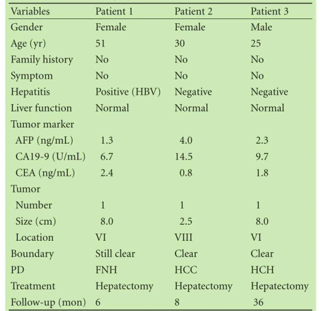
Table 1. Demographics and clinical characteristics
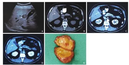
Fig. 1. Images and specimens of hepatic PEComas. A: ultrasonography of PEComa (arrow); B (plain CT scan) (arrow), C (arterial phase) (arrow), and D (venous phase): CT scan of hepatic PEComa (arrow); E: Operative specimens of hepatic PEComas (arrow).
Histologic analysis
The tumors were completely resected in the three patients. The diameters of the tumors were approximately 8 cm, 2.5 cm, and 8 cm, respectively. The cut surface was soft, and colored from yellow to dark red (Fig. 1E). The tumor specimens were evaluated by HE and IHC analysis. HE staining showed that the tumor was composed of thick-walled blood vessels, irregularly-arranged bundles of smooth muscle, and mature adipose tissue (Fig. 2). Immunohistochemically (Table 3), the tumor cells were strongly positive for HMB-45, Melan-A, and SMA in all patients (Table 3). Patient 1 was positive for E-cad, CD34, and Ki-67, patient 2 was positive for S-100 and Ki-67, and patient 3 was positive for E-cad and CD34. Tumor cells were negative for Desmin, Hep-1, CK8/18, CK7, and AFP.
Treatment and follow-up
All of the patients underwent hepatectomy. Intraoperative exploration for patient 1 revealed a soft mass with a rich blood supply, which was located between Couinaud VI and I. A rapid pathologic examination (RPE) showed a benign lesion with cloudy swelling of liver cells, interstitially-scattered inflammatory cell infiltration, no masses, and no nodules. Partial hepatectomy and selective hepatic artery ligation were performed. The postoperative IHC pathologic assessment was consistent with the diagnosis of a hepatic PEComa. Unfortunately, a
recurrence was detected 3 months postoperatively, and a second operation was performed in another hospital. No recurrence or metastasis was observed, and liver function test was normal during the follow-up for 3 months. Intraoperative exploration for patient 2 showed that a mass was located in Couinaud VIII between the right and middle hepatic veins. RPE suggested a benign lesion, and partial hepatectomy was performed. After an 8-month follow-up, no recurrence or metastasis was noted in this patient, who was finally lost to follow-up. Intraoperative exploration for patient 3 showed an exogenous mass located in Couinaud VI, and the result of RPE was consistent with a PEComa. Thus, radical hepatectomy was performed, and no tumor recurrence or metastasis occurred.
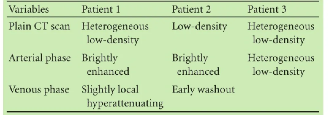
Table 2. Computed tomography of hepatic PEComas

Table 3. Immunohistochemistry of hepatic PEComas

Fig. 2. HE staining and immunohistology features of the PEComa. A: HE staining; B: CD34 staining; C: CK staining; D: HMB-45 staining; E: Melan-A staining; F: SMA staining.
Discussion
PEComa is a class of mesenchymal neoplasms consisting of three components: thick-walled, often hyalinized blood vessels; smooth muscle cells; and adipose tissue. PEComas often occur in the kidneys, lungs, pancreas, uterus, liver, etc.[5]The majority of hepatic PEComas are single, soft tissues, and solitary with a rich blood supply. PEComas have similar IHC profiles, including positivity for melanocytic (HMB-45) and SMA markers. Additional IHC markers such as S-100, CD34, and CD117 are helpful for confirming the diagnosis, and are not completely coincidental.[4,6,8]
Hepatic PEComas often occur in young women, and are commonly diagnosed in patients with abdominal pain or masses, occasionally as an incidental finding. The size of the lesion is usually relatively large (>5 cm), the largest lesion being 23×16×6 cm.[5]Most hepatic PEComas do not present characteristic images, which make the clinical diagnosis difficult. Recently, Liu’s group summarized the characteristic imaging findings of PEComas in five patients.[5]The tumors exhibited a distinctive mass with heterogeneous density on plain CT scans, and heterogeneous-enhancment in the arterial phase and an isoattenuating state in the portal venous phase. On magnetic resonance imaging (MRI), heterogeneous hypointensity was demonstrated on T1-weighted images (T1WIs) and hyperintensity on T2-weighted images (T2WIs). After fat saturation, the hyperintensity on T2WI in the liver lesion remained and heterogeneous enhancement appeared on arterial phase images. In our patients, a low density was shown on plain CT scans, bright or heterogeneous enhancement in the arterial phase, and early washout with a heterogeneous or isopycnic enhancement in the portal venous phase. These findings are different from hepatic cancers, most of which present with bright enhancement in the arterial phase and early washout with a lower density in the portal venous phase. In addition, hepatic focal nodular hyperplasia presents with enhancement in the portal venous phase.
The majority of PEComas are benign, whereas few malignant PEComas have been reported at present (Table 4).[9-17]Nguyen et al[18]suggested a differential diagnosis between benign and malignant hepatic PEComas: cytologic atypia; coagulative necrosis; large tumor size (>10 cm); negativity for CD117; and clinical evidence of aggressive disease. In the literature, myoid cells were reported positive for CD34[19]and Ki-67 in hepatic PEComas. In fact, we were the first to report that CD34 and Ki-67 were positive in a patient (patient 1). It is worth noting that the tumor recurred in patient 1 3 months after operation. CD34 and Ki-67 positivity indicates malignant behavior of hepatic PEComas. In addition, age, as prognostic marker of thyroid cancer, may be an important factor for identifying the biological characteristics of hepatic PEComas. The malignant hepatic PEComas reported in the literature (Table 4) mostly occurred in older (>50 years of age) women. In our patients, patient 1 with a malignant hepatic PEComa was a 51-year-old woman. Thus, hepatic PEComas may have a predilection for malignant behavior in older (>50 years of age) women. Taking into account the above factors, malignant hepatic PEComas should be considered in patients with the following characteristics: older (>50 years of age) females; abdominal discomfort and pain; larger tumor size
(>10 cm); positivity for CD34 and Ki-67; and negativity for CD117.
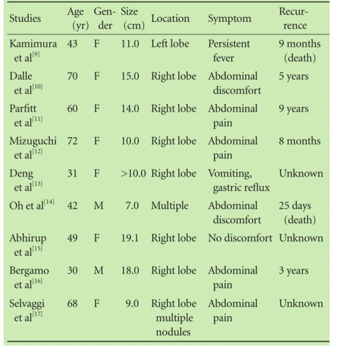
Table 4. Cases of malignant hepatic PEComas reported in the literature
Hepatectomy is the preferred therapeutic method for the treatment of PEComas, regardless of benign or malignant features. Surgical resection is feasible for the majority of patients. In fact, because of the rarity, malignant nature, and uncertainty of differential diagnosis (especially atypical hepatic cancer), hepatectomy is the best choice. In our patients, the tumor lesions were larger with cloudy swelling and no clear masses and nodules, thus microwave therapy was not feasible. According to a recent report, sirolimus, which suppresses mTOR signaling, may decrease the size of angiomyolipomas and improves lung function.[20]Sirolimus may be useful for the treatment of hepatic PEComa; however, at the presence of malignant behavior of hepatic PEComas, patients should be closely followed up after hepatectomy.
In conclusion, there is no validated method for the diagnosis of hepatic PEComas at present. Hepatic PEComas are borderline tumors, and may exhibit malignant behaviors in older (>50 years old) women accompanied with abdominal discomfort or pain, larger tumor size (>10 cm), positivity for CD34 and Ki-67, and negativity for CD117. Surgical resection is the preferred method, no matter whether the tumor is benign or malignant. Patients should be closely followed up after the resection.
Contributors: HBB and RJH contributed equally to this work. HBB, RJH and ZF proposed the study. RJH and ZF contributed to conception and design. HBB, FY, ZCY, and DXZ analyzed and interpreted the data; LX and LY collected the data; HBB and RJH wrote the article; RJH and ZF made the critical revision of the article. ZF is the guarantor.
Funding: This study was supported by grants from the Foundation of Jiangsu Collaborative Innovation Center of Biomedical Functional Materials, Basic Research Program-Youth Fund Project of Jiangsu Province (BK20140092), and the National Natural Science Foundation of China (81400650, 81273261 and 81270583). Ethical approval: Not needed.
Competing interest: No benefits in any form have been received or will be received from a commercial party related directly or indirectly to the subject of this article.
1 Folpe AL. Neoplasms with perivascular epithelioid cell differentiation (PEComas). In Pathology and Genetics of Tumours of Soft Tissue and Bone. Edited by Fletcher CDM, Unni KK, Epstein J, Mertens F. Lyon: IARC Press; 2002:221-222.
2 Akitake R, Kimura H, Sekoguchi S, Nakamura H, Seno H, Chiba T, et al. Perivascular epithelioid cell tumor (PEComa) of the liver diagnosed by contrast-enhanced ultrasonography. Intern Med 2009;48:2083-2086.
3 Fang SH, Zhou LN, Jin M, Hu JB. Perivascular epithelioid cell tumor of the liver: a report of two cases and review of the literature. World J Gastroenterol 2007;13:5537-5539.
4 Paiva CE, Moraes Neto FA, Agaimy A, Custodio Domingues MA, Rogatto SR. Perivascular epithelioid cell tumor of the liver coexisting with a gastrointestinal stromal tumor. World J Gastroenterol 2008;14:800-802.
5 Liu Z, Qi Y, Wang C, Zhang X, Wang B. Hepatic perivascular epithelioid cell tumor: five case reports and literature review. Asian J Surg 2015;38:58-63.
6 Petrolla AA, Xin W. Hepatic angiomyolipoma. Arch Pathol Lab Med 2008;132:1679-1682.
7 Neofytou K, Famularo S, Khan AZ. PEComa in a young patient with known Li-Fraumeni syndrome. Case Rep Med 2015;2015: 906981.
8 Li T, Wang L, Yu HH, Sun HC, Qin LX, Ye QH, et al. Hepatic angiomyolipoma: a retrospective study of 25 cases. Surg Today 2008;38:529-535.
9 Kamimura K, Oosaki A, Sugahara S, Mori S, Moroda T, Satoh O, et al. Malignant potential of hepatic angiomyolipoma: case report and literature review. Clin J Gastroenterol 2010;3:104-110.
10 Dalle I, Sciot R, de Vos R, Aerts R, van Damme B, Desmet V, et al. Malignant angiomyolipoma of the liver: a hitherto unreported variant. Histopathology 2000;36:443-450.
11 Parfitt JR, Bella AJ, Izawa JI, Wehrli BM. Malignant neoplasm of perivascular epithelioid cells of the liver. Arch Pathol Lab Med 2006;130:1219-1222.
12 Mizuguchi T, Katsuramaki T, Nobuoka T, Nishikage A, Oshima H, Kawasaki H, et al. Growth of hepatic angiomyolipoma indicating malignant potential. J Gastroenterol Hepatol 2004;19:1328-1330.
13 Deng YF, Lin Q, Zhang SH, Ling YM, He JK, Chen XF. Malignant angiomyolipoma in the liver: a case report with pathological and molecular analysis. Pathol Res Pract 2008;204:911-918.
14 Oh HW, Kim TH, Cha RR, Kim NY, Kim HJ, Jung WT, et al. A case of malignant perivascular epithelioid cell tumor of the retroperitoneum with multiple metastases. Korean J Gastroenterol 2014;64:302-306.
15 Abhirup B, Kaushal K, Sanket M, Ganesh N. Malignant hepatic perivascular epithelioid cell tumor (PEComa) - case report and a brief review. J Egypt Natl Canc Inst 2015;27:239-242.
16 Bergamo F, Maruzzo M, Basso U, Montesco MC, Zagonel V, Gringeri E, et al. Neoadjuvant sirolimus for a large hepatic perivascular epithelioid cell tumor (PEComa). World J Surg Oncol 2014;12:46.
17 Selvaggi F, Risio D, Claudi R, Cianci R, Angelucci D, Pulcini D, et al. Malignant PEComa: a case report with emphasis on clinical and morphological criteria. BMC Surg 2011;11:3.
18 Nguyen TT, Gorman B, Shields D, Goodman Z. Malignant hepatic angiomyolipoma: report of a case and review of literature. Am J Surg Pathol 2008;32:793-798.
19 Sturtz CL, Dabbs DJ. Angiomyolipomas: the nature and expression of the HMB45 antigen. Mod Pathol 1994;7:842-845.
20 Bissler JJ, McCormack FX, Young LR, Elwing JM, Chuck G, Leonard JM, et al. Sirolimus for angiomyolipoma in tuberous sclerosis complex or lymphangioleiomyomatosis. N Engl J Med 2008;358:140-151.
Received September 2, 2015
Accepted after revision December 23, 2015
(Hepatobiliary Pancreat Dis Int 2016;15:660-664)
Author Affiliations: Department of Liver Surgery (Hao BB, Rao JH, Fan Y, Zhang CY, Dai XZ and Zhang F) and Department of Pathology (Li X and Leng Y), First Affiliated Hospital of Nanjing Medical University; Key Laboratory of Living Donor Liver Transplantation of Ministry of Public Health, Nanjing 210029, China
Feng Zhang, MD, PhD, Department of Liver Surgery, First Affiliated Hospital of Nanjing Medical University; Key Laboratory of Living Donor Liver Transplantation of Ministry of Public Health, 300 Guangzhou Road, Nanjing 210029, China (Tel: +86-25-83718836 ext 6476; Fax: +86-25-83672106; Email: zhangfeng1958@hotmail.com)
? 2016, Hepatobiliary Pancreat Dis Int. All rights reserved.
10.1016/S1499-3872(16)60077-2
Published online March 2, 2016.
 Hepatobiliary & Pancreatic Diseases International2016年6期
- Hepatobiliary & Pancreatic Diseases International的其它文章
- Postoperative day one serum alanine aminotransferase does not predict patient morbidity and mortality after elective liver resection in non-cirrhotic patients
- Uncoupling protein 2 deficiency reduces proliferative capacity of murine pancreatic stellate cells
- Jagged1 and DLL4 expressions in benign and malignant pancreatic lesions and their clinicopathological significance
- Endoscopic bilateral stent-in-stent placement for malignant hilar obstruction using a large cell type stent
- Prognosis of hepatocellular carcinoma with bile duct tumor thrombus after R0 resection: a matched study
- Urgent ERCP for acute cholangitis reduces mortality and hospital stay in elderly and very elderly patients