
Continuous Flow Left Ventricular Assist Device Therapy: A Focused Review on Optimal Patient Selection and Long-Term Follow-up Using Echocardiography
2015-05-22 03:33:40JuanVilaroMDAnitaSzadyMDMustafaAhmedMDJacquelineDawsonMDandJuanArandaJrMD
Juan R. Vilaro, MD, Anita Szady, MD, Mustafa M. Ahmed, MD, Jacqueline Dawson, MD and Juan M. Aranda Jr., MD
1North Florida/South Georgia Veterans Health System, Cardiology Section, Gainesville, FL, USA
2University of Florida College of Medicine, Division of Cardiovascular Medicine, Gainesville, FL, USA
3Western Kentucky Heart and Lung Associates, Division of Cardiology, Scottsville, KY, USA
Introduction
Despite widespread awareness and use of scientif i cally proven life-prolonging medical and device-based therapies over the last two decades,heart failure remains a leading cause of morbidity, mortality, and health care expenditure in the United States [1]. Patients who have progressive heart failure syndromes despite receiving standard treatment, including medical therapy and cardiac resynchronization, are being increasingly considered for implantation of continuous-f l ow left ventricular (LV) assist devices (CF-LVADs). CFLVADs are effective in improving survival and quality of life in patients with advanced heart failure, and can be used as destination therapy or as a bridge to heart transplantation [2, 3]. The increasing incidence of CF-LVAD implantation over the last decade mandates an effort from all practicing physicians, not just heart failure specialists,to understand the basic anatomic and physiologic implications of these devices on cardiac structure and function. Echocardiography plays an integral role in the evaluation of patients with advanced heart failure who are being considered for or are mechanically supported by a CF-LVAD. Here we provide a focused clinical review on the use of echocardiography in two main aspects of the evaluation of these patients: (a) optimal patient selection for CF-LVAD support and (b) follow-up assessment of optimal pump function.
Optimal Patient Selection
In patients being considered for durable CF-LVAD implantation, the preoperative echocardiogram is a critical part of the evaluation and can be a powerful predictor of short-term and long-term clinical outcomes after implantation. The preoperative echocardiogram should focus on the following:
· Detailed assessment of right ventricular (RV)and LV size, structure, and function
· Presence of signif i cant valvular regurgitation,particularly of the aortic and tricuspid valves
· Presence of intracardiac thrombi
· Presence of intracardiac shunts
Left Ventricular Structure and Function
Patients being considered for CF-LVAD placement almost invariably have severe reduction in their LV systolic function. Consideration of mechanical circulatory support, including CF-LVADs, is not recommended if the LV ejection fraction is greater than 25% [4]. Careful measurement of LV dimensions is also important, as the presence of relatively small LV cavities, specifically smaller than 63 mm, has been independently associated with increased morbidity and mortality after CF-LVAD implantation [5]. This is likely related to excessive LV emptying and exaggerated leftward shift of the interventricular septum due to its close proximity to the inf l ow cannula. This abnormal septal deformation worsens RV function [6], and can also cause direct physical contact of the septum with the inf l ow cannula, termed a suction event. Suction events prevent optimal function of the CF-LVAD as they automatically trigger speed decrements and are frequently associated with ventricular arrhythmias [7]. From a structural and functional standpoint, hearts with a severely reduced LV ejection fraction and moderate to severe degrees of LV dilation are therefore likeliest to benef i t from CF-LVAD support.
Right Ventricular Structure and Function
Assessment of RV function is critical in patients being considered for LV assist device (LVAD)implantation. The hemodynamic effects of a CFLVAD on the right ventricle can be divergent. The desired favorable effect is improvement in RV performance following unloading of the left side of the heart and subsequent decongestion of the pulmonary circulation. However, several hemodynamic consequences on the right ventricle following initiation of CF-LVAD support challenge the right side of the heart. Three potentially detrimental effects on right-sided heart function are (1) acute increase in right ventricular preload (which requires an equivalent increase in right-sided cardiac output),(2) leftward septal deformation resulting from LV unloading leading to worsening RV function, and(3) worsening tricuspid regurgitation.
There are numerous validated echocardiographic measures of RV structure and function [8]. This review will focus on two indices that have been studied in CF-LVAD patients, are reproducible, and are easily obtainable: (a) tricuspid annular plane systolic excursion (TAPSE) and (b) heart rate– corrected duration of tricuspid regurgitation (TRDc).
TAPSE represents the distance of longitudinal motion of the tricuspid annulus during systole and is a validated index of RV systolic function [8]. It is obtained by M-mode imaging of the lateral tricuspid annulus obtained from an apical four-chamber view. An example is illustrated in Figure 1. TAPSE values below 7.5 mm have high specificity for postoperative right-sided heart failure, and may indicate a need for more aggressive preoperative hemodynamic optimization and/or the need for prolonged inotropic support following CF-LVAD implantation [9].
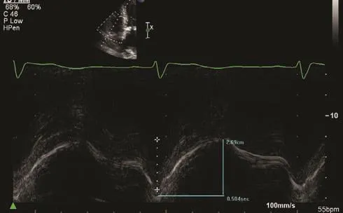
Figure 1 Assessment of Right Ventricular Systolic Function by Tricuspid Annular Plane Systolic Excursion (TAPSE) Measurement. The Longitudinal Motion of the Lateral Tricuspid Annulus is 27 mm, Consistent with Normal Right Ventricular Systolic Function (Normal is More Than 16 mm).
TRDc is a novel index described by Topilsky et al. [5] in a large cohort of CF-LVAD patients from the Mayo Clinic. One calculates it by measuring the duration of the tricuspid regurgitant Doppler jet and dividing it by the square root of the R-R interval. TRDc is a heart rate–adjusted measure of the time to pressure equalization between the right ventricle and the right atrium during systole that integrates right atrial compliance and the severity of tricuspid regurgitation. In this regard, it can predict the hemodynamic impact of an acute volume load increase on the right atrium, an expected result of CF-LVAD implantation. Not surprisingly, it was found on multivariate analysis to have a strong ability to predict rates of RV failure and death following LVAD implantation. Patients with a TRDc of less than 461 ms had an adjusted 2-year mortality odds ratio of 2.3 compared with patients with a TRDc longer than 461 ms. Examples of TRDc that would predict low and high risks of RV failure and death are illustrated in Figure 2.
Valvular Insuffi ciency
Accurate assessment of any underlying valvular regurgitation, particularly of the aortic and tricuspid valves, is crucial in the evaluation of patients being considered for CF-LVADs, as it directly impacts decisions to perform additional surgery at the time of implantation.
Aortic Regurgitation
There are several validated quantitative and qualitative methods for assessing the severity of aortic insuff i ciency (AI) [10]. Quantitative methods include regurgitant orif i ce and regurgitant volume calculation, which can be done with spectral Doppler imaging or the proximal isovelocity surface area method, and vena contracta or jet width measurement. Qualitative measures include pressure half-time and degree of descending aorta diastolic fl ow reversal. A brief summary of quantitative and qualitative estimates of AI severity is given in Table 1. It is important to remember that in patients with advanced heart failure, LV filling pressures are almost invariably elevated and the pressure halftime method may overestimate the severity of AI signifi cantly [11]. The severity of any preexisting AI will typically worsen to some degree following initiation of CF-LVAD support [12]. Although CFLVAD support is in many cases well tolerated in patients with minimal or mild AI, the presence of moderate AI or worse can result in a physiologically ineffective circuit (left ventricle → inf l ow cannula→ outf l ow cannula → ascending aorta → left ventricle) owing to a large fraction of the blood volume exiting the outf l ow graft regurgitating into the left ventricle [13]. For this reason, any patient with AI of more than mild severity should also undergo concomitant aortic valve closure at the time of LVAD implantation. Repair with a single coaptation stitch at the time of implantation provides effective and durable repair of moderate or severe AI in patients in whom a CF-LVAD has been implanted [14].
Tricuspid Regurgitation
A detailed assessment of tricuspid valve structure and function is key in the preoperative assessment of patients being considered for CF-LVAD implantation, particularly when there is any signif i cant degree of tricuspid regurgitation. Similarly to its effects on overall RV function, the hemodynamic effects of CF-LVAD support on tricuspid regurgitation may result in worsening, unchanged, or improved tricuspid regurgitation severity [15, 16].The acute increase in RV end-diastolic volumes following CF-LVAD support can cause functional tricuspid regurgitation to worsen, leading to ineffective forward fl ow and a syndrome of progressive RV failure. The severity of tricuspid regurgitation at the baseline is associated with increased risk of RV failure when it is moderate or worse on the preoperative transthoracic echocardiogram [17].However, there are conf l icting data regarding the benef i ts of tricuspid valve repair at the time of CFLVAD implantation in patients with moderate or severe tricuspid regurgitation, and the decision to perform concomitant tricuspid valve repair is ultimately deferred to the performing surgeon [18–20].Tricuspid regurgitation severity can be graded quantitatively by vena contracta width or tricuspid regurgitant jet area, or qualitatively by evaluation of the Doppler prof i le of the hepatic vein (systolic fl ow reversal implies severe tricuspid regurgitation)[10]. An illustration of severe tricuspid regurgitation by color Doppler imaging as well as hepatic vein systolic fl ow reversal is illustrated in Figure 3.Table 2 brief l y summarizes the parameters of tricuspid regurgitation severity.
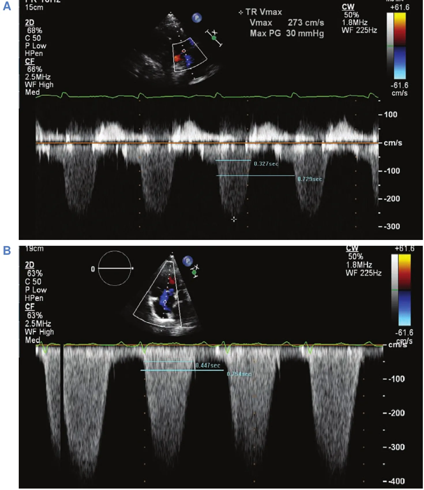
Figure 2 (A) Example of a Patient with a Heart Rate–corrected Tricuspid Regurgitation Duration (TRDc) Conferring a High Risk of Postoperative Right-sided Heart Failure and Death (TRDc = 384 ms). (B) Example of a patient with a TRDc conferring a low risk of right ventricular failure and death (TRDc = 519 ms).
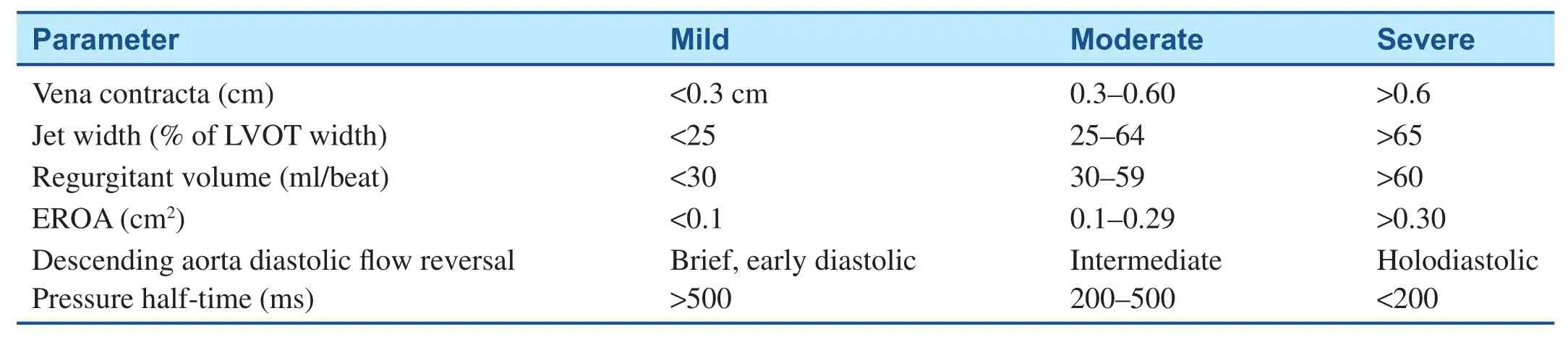
Table 1 Echocardiographic Parameters Used in Grading the Severity of Aortic Regurgitation.
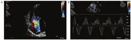
Figure 3 (A) Example of severe tricuspid regurgitation by color Doppler imaging. Note the broad-based regurgitant jet reaching the posterior wall of the right atrium. (B) Pulsed-wave Doppler imaging demonstrating hepatic vein systolic fl ow reversal,consistent with severe tricuspid regurgitation.
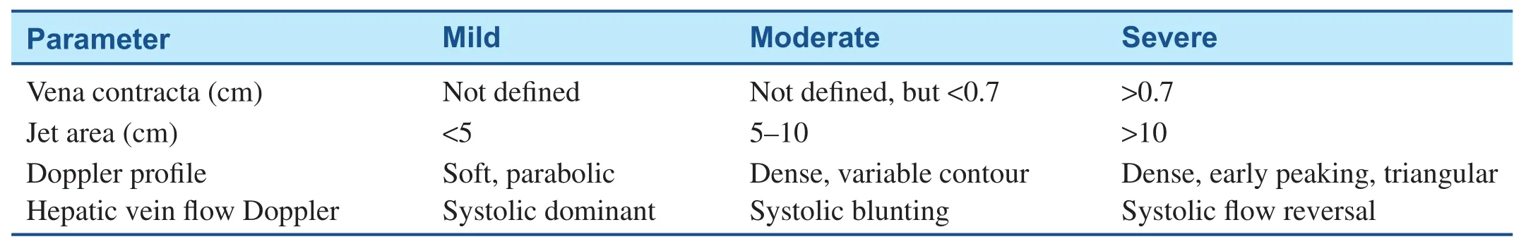
Table 2 Echocardiographic Parameters Used in Grading the Severity of Tricuspid Regurgitation.
Evaluation for Intracardiac Thrombi
Preoperative identif i cation of intracardiac thrombus, particularly in the LV apex, which is not uncommon with dilated cardiomyopathy and a severely reduced LV ejection fraction, is also an important part of preoperative planning before CFLVAD implantation. Although the presence of an apical thrombus does not contraindicate placement of a CF-LVAD, it requires removal of the thrombus at the time of surgery before insertion of the inf l ow cannula in the LV apex. One study reported that of 100 patients in whom an LVAD has been implanted over 3 years, six had an LV apical thrombus identif i ed preoperatively or intraoperatively.None of them experienced a neurological event,pump thrombosis, or pump malfunction [21]. LV thrombi can be readily detected by transthoracic echocardiography, typically in the apical views. If images are of limited quality, echocardiography contrast agents should be used as they signifi cantly improve the sensitivity, specificity, and accuracy of echocardiography in diagnosing LV thrombus [22].An example is shown in Figure 4.
Presence of Intracardiac Shunts and Patent Foramen Ovale
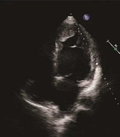
Figure 4 Apical Four-Chamber View Demonstrating Mural Left Ventricular Apical Thrombus in a Patient with Recent Anterior Transmural Myocardial Infarction.
The echocardiogram before CF-LVAD implantation should include careful inspection for any evidence of intracardiac shunting, including a patent foramen ovale (PFO). A PFO are not uncommon and has a reported prevalence of up to 25% in the general population [23]. Although it is typically noted incidentally and not felt to cause any signif i cant shunting,the acute lowering of left-sided intracardiac pressures resulting from CF-LVAD support can precipitate increased right to left interatrial shunting and clinically important hypoxemia and cyanosis [24].Preoperatively, transthoracic echocardiography can identify atrial level communication with color Doppler imaging of the interatrial septum, or in the apical windows following intravenous administration of agitated saline [25]. The appearance of agitated saline bubbles in the left heart chambers within three beats or less of their appearance in the right side of the heart is typically felt to represent the presence of an intracardiac shunt, most commonly a PFO. It is, however, important to remember that in patients with advanced heart failure and signifi cantly elevated right-and left-sided atrial pressures there may not be a high enough gradient between both atria to cause a detectable shunt. Therefore, in addition to preoperative inspection for shunting, the intraoperative transesophageal echocardiogram should be used to confirm the presence or absence of any shunting,including a PFO that may not have been detected by transthoracic echocardiography. A PFO identif i ed in patients undergoing CF-LVAD implantation should be closed at the time of surgery to eliminate the risk of hypoxemia from right to left shunting.
Follow-up Echocardiographic Assessment After Continuous-Flow Left Ventricular Assist Device Placement
After surgical implantation of a CF-LVAD, echocardiography continues to be an essential clinical tool for the ongoing evaluation and follow-up of pump function, native cardiac function, and overall patient care. The key elements of an echocardiogram in the patient supported with a CF-LVAD should focus on the following:
· Evaluation of adequate LV unloading:
· Frequency of aortic valve opening
· LV dimension and interventricular septal morphology
· Inf l ow and outf l ow cannula velocities
· Detailed assessment of RV function, including serial assessment over time
Evaluating Adequate Left Ventricular Unloading in Patients Supported by a Continuous-Flow Left Ventricular Assist Device
There are multiple features of the echocardiogram that can provide evidence of adequate LV unloading,which is invariably the primary hemodynamic goal of CF-LVAD support. These include the degree and frequency of aortic valve opening, the change in LV dimensions over time, and the cannula velocities.
Aortic Valve Opening
Evaluation of aortic valve opening by echocardiography is a simple, reliable way of determining adequate LV unloading in patients following CF-LVAD implantation [26]. In patients with CF-LVAD support, the aortic valve typically should open not every beat, but rather intermittently every two to three beats, or not at all. The frequency of aortic valve opening can be assessed by 2D imaging in the parasternal long-axis and short-axis views, as well as the apical three-chamber view. However, postsurgically image quality is frequently limited, and M-mode imaging through the aortic valve plane can assess the degree of leaf l et opening and excursion more def i nitively (Figure 5). An aortic valve that opens consistently with each systole suggests that the left ventricle is not adequately unloaded and LV pressures remain signifi cantly elevated. Common causes of inadequate LV unloading include uncontrolled hypertension, intravascular hypervolemia,any form of mechanical obstruction in the pump,including thrombus or kinking of the outf l ow graft,and the speed setting being too low. Table 3 summarizes the different clinical features of the most common causes of poor LV unloading.
Septal Morphology and Ventricular Dimensions
Figure 5 M-mode Image of the Aortic Valve from Patient with Continuous-f l ow Left Ventricular Assist Device Support.Note the minimal aortic leaf l et excursion indicative of adequate unloading of the left ventricle.
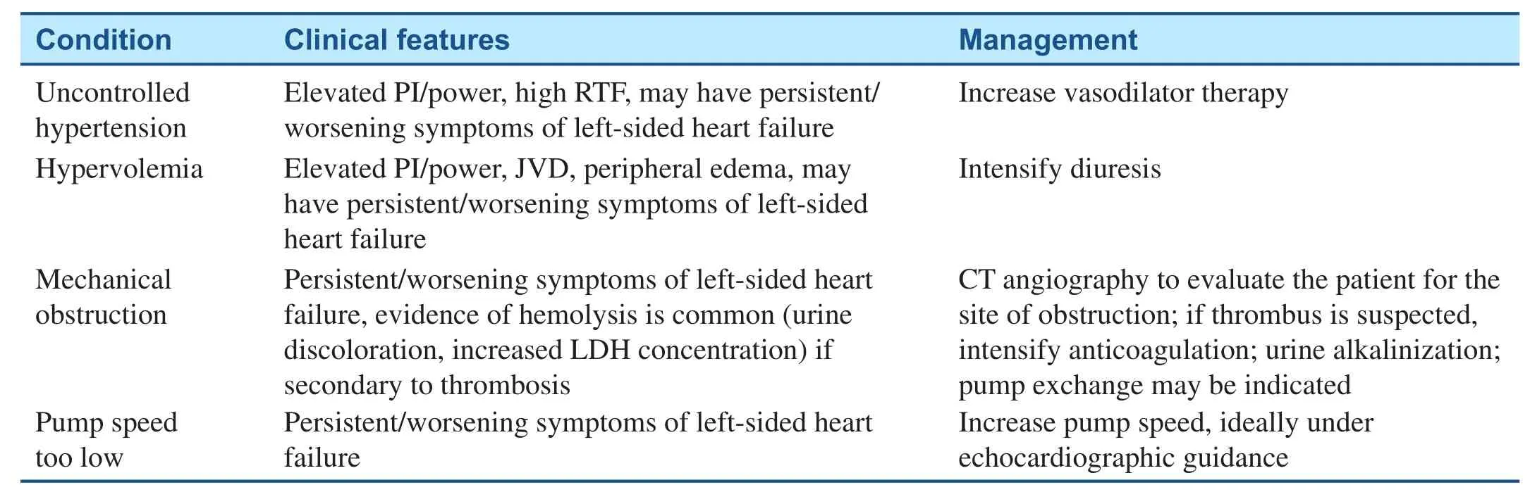
Table 3 Causes of Poor Left Ventricular Unloading While the Patient has Continuous-f l ow Left Ventricular Assist Device Support.
Follow-up echocardiographic assessment of LV chamber dimensions, together with the morphology and shape of the interventricular septum, can also provide information regarding the adequacy of LV unloading. Following initiation of CF-LVAD support, LV dimensions typically decrease and slight leftward motion of the interventricular septum is expected, ref l ecting a moderate decrease in LV pressures relative to RV pressures [27]. If, however,there is an interval increase of the LV dimension, or if there is a predominantly rightward motion of the septum, this suggests increased LV pressure relative to RV pressure, and is typically a sign of inadequate LV unloading. The differential is similar to that of an aortic valve that opens constantly, and workup for all of the previously mentioned scenarios should be pursued (Table 3).
Conversely, if the leftward septal shift is pronounced or associated with a dramatic reduction in LV cavity size, this highly suggests excessive LV decompression by the pump. Common causes of this include the pump speed being too high, signif i -cant recovery of LV systolic function, or any scenario of reduced LV preload, including intravascular hypovolemia or right-sided heart failure. Table 4 summarizes a diagnostic approach to help distinguish between these different scenarios, as well as potential therapeutic options.
A ramp study should be considered whenever there is clinical evidence of poor LV unloading due to suspected mechanical obstruction [28]. This consists in measuring echocardiographic LV dimensions and the frequency of aortic valve opening while serially increasing the CF-LVAD pump speed. An inability to decrease LV dimensions or reduce the frequency of aortic valve opening despite increasing pump speeds should raise concern for mechanical obstruction in the pump, including pump thrombosis.
Cannula Velocities
The echocardiogram in patients supported with CFLVADs should include attempts to measure blood fl ow velocities through the inf l ow cannula and outf l ow graft. However, Doppler measurements of inf l ow and outf l ow velocities are rarely interpretable in centrifugal-f l ow CF-LVADs because of multiple artifacts [29]. In axial-f l ow LVADs, inf l ow velocities are best evaluated in the apical windows,where fl ow is most parallel to the ultrasound beam.Outf l ow graft velocities can be measured in the parasternal long-axis window or the suprasternal window as blood fl ows out of the pump into the ascending aorta. There is no consensus on what the normal range of cannula or graft velocities should be. Ideally, there should be laminar fl ow by color Doppler imaging, with minimal turbulence, which suggests adequate alignment with mitral inf l ow. By spectral Doppler imaging, velocities vary widely depending on the patient and loading conditions,typically ranging between 0.3 and 1.5 m/s [30].Most patients have some degree of phasic variationin velocity over the cardiac cycle, which ref l ects changes in fl ow through the pump due to native ventricular contraction and diastolic mitral inf l ow(Figure 6). Although there is not an establishednormalrange of inf l ow cannula or outf l ow graft velocities, any signif i cant change in these velocities over time warrants further evaluation and should be interpreted in the clinical context of the individual patient.
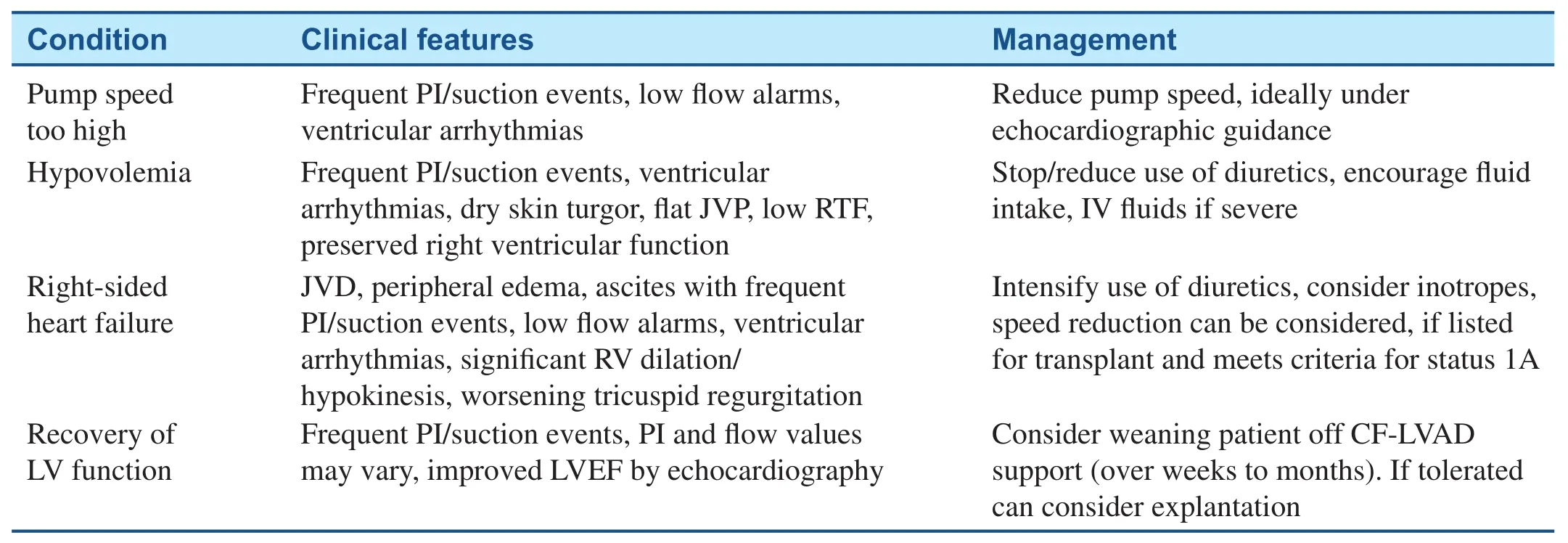
Table 4 Causes of Excessive Left Ventricular Decompression.
Assessment of Right Ventricular Function
As discussed previously, it is crucial for patients supported by CF-LVADs to have relatively preserved RV function in order to maintain adequate rightsided cardiac output. Syndromes of right-sided heart failure can occur acutely in the early postoperative period or can have a more chronic presentation several years after implantation [31, 32]. Any interval worsening of RV dilation or the previously described indices of RV function support the diagnosis of RV failure after CF-LVAD implantation in the appropriate clinical context. In addition, echocardiography also provides a simple noninvasive way of detecting hemodynamics suggestive of RV decompensation.
Patients with right-sided heart failure after ventricular assist device (VAD) implantation typically experience a syndrome of RV volume overload as the right side of the heart struggles to match the effective forward fl ow provided by a CF-LVAD.As this progresses, the right ventricle becomes progressively dilated, functional tricuspid regurgitation worsens, and right-sided filling pressures become severely elevated. The echocardiographic estimation of right atrial and RV systolic pressure using the inferior vena cava diameter and tricuspid regurgitant jet velocity are well validated [8]. These methods appear to retain their accuracy in patients following CF-LVAD implantation [33]. Signif i cant dilation of the inferior vena cava with minimal or no inspiratory collapse together with a relatively low tricuspid regurgitant jet velocity (less than 2.5 m/s in the setting of a dilated inferior vena cava) is the echocardiographic correlate of RV failure diagnosed by invasive hemodynamics, and highly suggests post–VAD implantation right-sided heart failure. The onset of RV failure after VAD implantation portends a poor prognosis and increased mortality.Treatment options are limited unless patients are candidates for heart transplantation or biventricular support [31]. Symptomatically, patients benef i t from intravenous diuretics and initiation of inotropes for RV support. If septal morphology and ventricular chamber dimensions suggest excessive LV emptying and severe asymmetric dilation of the right ventricle relative to the left ventricle, a decrease in pump speed can also be helpful. Figure 7 illustrates images from a patient with post–VAD implantation right-sided heart failure.
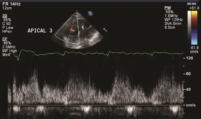
Figure 6 Inf l ow Cannula Velocity Measured from an Off-axis Apical Three-chamber View from a Patient with a Normally Functioning Continuous-f l ow Left Ventricular Assist Device.
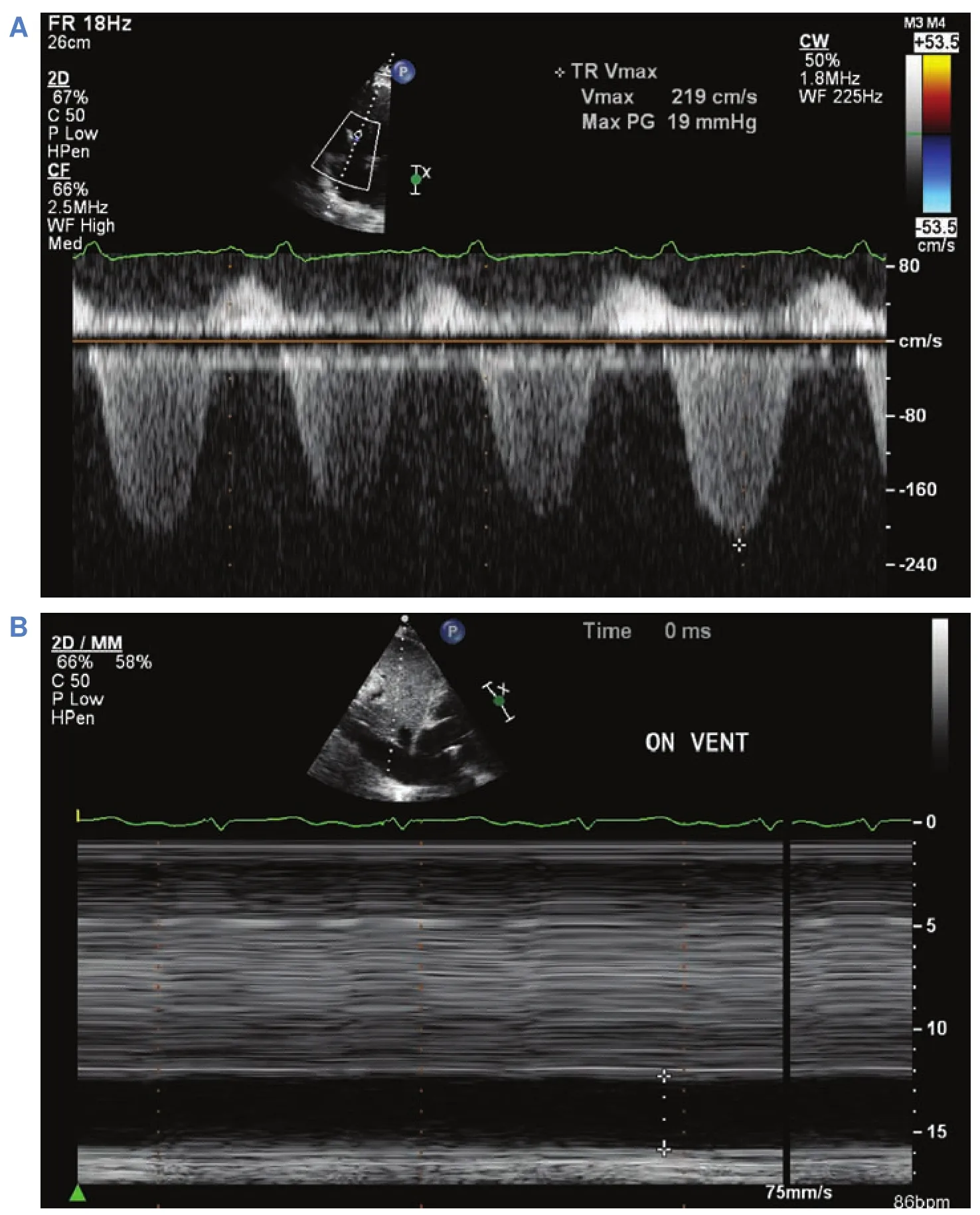
Figure 7 Tricuspid Regurgitation Doppler and Inferior Vena Cava M-mode Imaging from a Patient with Severe Right Ventricular Failure Following Continuous-f l ow Left Ventricular Assist Device Placement.
Conclusion and Take-Home Message
The incidence of advanced heart failure patients who are potential candidates for mechanical support with CF-LVADs is steadily increasing worldwide. Echocardiography is a simple, noninvasive, yet highly useful diagnostic imaging modality that provides easily interpretable information that is instrumental in the care of patients supported by this relatively complex technology. It is crucial to remember that the utility of echocardiography is best when the images are understood in the clinical context of each individual patient, and decisions should never be made solely on the basis of the results of a single study.
Conflict of Interest
The authors declare no conf l ict of interest.
Funding
This research received no specific grant from any funding agency in the public, commercial or notfor-profit sectors.
1. Fonarow GC, Yancy CW,Hernandez AF, Peterson ED,Spertus JA, Heidenreich PA. Potential impact of optimal implementation of evidence-based heart failure therapies on mortality. Am Heart J 2011;161:1024–30.e1023.
2. Christiansen S, Brose S,Autschbach R. [Surgical therapy of end-stage heart failure]. Herz 2003;28:380–92.
3. Miller LW, Pagani FD, Russell SD, John R, Boyle AJ, Aaronson KD, et al. Use of a continuousflow device in patients awaiting heart transplantation. N Engl J Med 2007;357:885–96.
4. Yancy CW, Jessup M, Bozkurt B,Butler J, Casey DE, Drazner MH,et al. 2013 ACCF/AHA guideline for the management of heart failure: a report of the American College of Cardiology Foundation/American Heart Association Task Force on Practice Guidelines. J Am Coll Cardiol 2013;62:e147–239.
5. Topilsky Y, Oh JK, Shah DK,Boilson BA, Schirger JA, Kushwaha SS, et al. Echocardiographic predictors of adverse outcomes after continuous left ventricular assist device implantation. JACC Cardiovasc Imaging 2011;4:211–22.
6. Neragi-Miandoab S, Goldstein D,Bello R, Michler R, D’Alessandro D. Right ventricular dysfunction following continuous fl ow left ventricular assist device placement in 51 patients: predicators and outcomes.J Cardiothorac Surg 2012;7:60.
7. Vollkron M, Voitl P, Ta J,Wieselthaler G, Schima H. Suction events during left ventricular support and ventricular arrhythmias. J Heart Lung Transplant 2007;26:819–25.
8. Rudski LG, Lai WW, Af i lalo J,Hua L, Handschumacher MD,Chandrasekaran K, et al. Guidelines for the echocardiographic assessment of the right heart in adults: a report from the American Society of Echocardiography endorsed by the European Association of Echocardiography, a registered branch of the European Society of Cardiology, and the Canadian Society of Echocardiography. J Am Soc Echocardiogr 2010;23:685–713;quiz 786-688.
9. Puwanant S, Hamilton KK, Klodell CT, Hill JA, Schof i eld RS, Cleeton TS, et al. Tricuspid annular motion as a predictor of severe right ventricular failure after left ventricular assist device implantation. J Heart Lung Transplant 2008;27:1102–7.
10. Zoghbi WA, Enriquez-Sarano M,Foster E, Grayburn PA, Kraft CD,Levine RA, et al. Recommendations for evaluation of the severity of native valvular regurgitation with two-dimensional and Doppler echocardiography. J Am Soc Echocardiogr 2003;16:777–802.
11. Griff i n BP, Flachskampf FA, Siu S, Weyman AE, Thomas JD. The effects of regurgitant orif i ce size,chamber compliance, and systemic vascular resistance on aortic regurgitant velocity slope and pressure half-time. Am Heart J 1991;122:1049–56.
12. Topilsky Y, Oh JK, Atchison FW,Shah DK, Bichara VM, Schirger JA, et al. Echocardiographic fi ndings in stable outpatients with properly functioning HeartMate II left ventricular assist devices. J Am Soc Echocardiogr 2011;24:157–69.
13. Gregory SD, Stevens MC, Wu E,Fraser JF, Timms D. In vitro evaluation of aortic insuff i ciency with a rotary left ventricular assist device.Artif Organs 2013;37:802–9.
14. McKellar SH, Deo S, Daly RC,Durham LA, Joyce LD, Stulak JM,et al. Durability of central aortic valve closure in patients with continuous fl ow left ventricular assist devices. J Thorac Cardiovasc Surg 2014;147:344–8.
15. Atluri P, Fairman AS, MacArthur JW, Goldstone AB, Cohen JE,Howard JL, et al. Continuous fl ow left ventricular assist device implant signifi cantly improves pulmonary hypertension, right ventricular contractility, and tricuspid valve competence. J Card Surg 2013;28:770–5.
16. Piacentino V, Williams ML, Depp T, Garcia-Huerta K, Blue L, Lodge AJ, et al. Impact of tricuspid valve regurgitation in patients treated with implantable left ventricular assist devices. Ann Thorac Surg 2011;91:1342–6; discussion 1346–7.
17. Potapov EV, Stepanenko A, Dandel M, Kukucka M, Lehmkuhl HB,Weng Y, et al. Tricuspid incompetence and geometry of the right ventricle as predictors of right ventricular function after implantation of a left ventricular assist device. J Heart Lung Transplant 2008;27:1275–81.
18. Piacentino V, Ganapathi AM,Stafford-Smith M, Hsieh MK,Patel CB, Simeone AA, et al. Utility of concomitant tricuspid valve procedures for patients undergoing implantation of a continuous-f l ow left ventricular device. J Thorac Cardiovasc Surg 2012;144:1217–21.
19. Maltais S, Topi lsky Y,Tchantchaleishvili V, McKellar SH, Durham LA, Joyce LD, et al.Surgical treatment of tricuspid valve insuff i ciency promotes early reverse remodeling in patients with axial-f l ow left ventricular assist devices. J Thorac Cardiovasc Surg 2012;143:1370–6.
20. Robertson JO, Grau-Sepulveda MV, Okada S, O’Brien SM,Matthew Brennan J, Shah AS, et al.Concomitant tricuspid valve surgery during implantation of continuous-f l ow left ventricular assist devices: a Society of Thoracic Surgeons database analysis. J Heart Lung Transplant 2014;33:609–17.
21. Engin C, Yagdi T, Balcioglu O,Erkul S, Baysal B, Oguz E, et al. Left ventricular assist device implantation in heart failure patients with a left ventricular thrombus. Transplant Proc 2013;45:1017–9.
22. Thanigaraj S, Schechtman KB,Pérez JE. Improved echocardiographic delineation of left ventricular thrombus with the use of intravenous second-generation contrast image enhancement. J Am Soc Echocardiogr 1999;12:1022–6.
23. Meissner I, Whisnant JP,Khandheria BK, Spittell PC,O’Fallon WM, Pascoe RD, et al.Prevalence of potential risk factors for stroke assessed by transesophageal echocardiography and carotid ultrasonography: the SPARC study.Stroke Prevention: Assessment of Risk in a Community. Mayo Clin Proc 1999;74:862–9.
24. Srinivas CV, Collins N, Borger MA, Horlick E, Murphy PM.Hypoxemia complicating LVAD insertion: novel application of the Amplatzer PFO occlusion device. J Card Surg 2007;22:156–8.
25. Marriott K, Manins V, Forshaw A,Wright J, Pascoe R. Detection of right-to-left atrial communication using agitated saline contrast imaging: experience with 1162 patients and recommendations for echocardiography. J Am Soc Echocardiogr 2013;26:96–102.
26. Estep JD, Stainback RF, Little SH,Torre G, Zoghbi WA. The role of echocardiography and other imaging modalities in patients with left ventricular assist devices. JACC Cardiovasc Imaging 2010;3:1049–64.
27. Liao KK, Miller L, Toher C,Ormaza S, Herrington CS, Bittner HB, et al. Timing of transesophageal echocardiography in diagnosing patent foramen ovale in patients supported with left ventricular assist device. Ann Thorac Surg 2003;75:1624–6.
28. Uriel N, Morrison KA, Garan AR, Kato TS, Yuzefpolskaya M,Latif F, et al. Development of a novel echocardiography ramp test for speed optimization and diagnosis of device thrombosis in continuous-f l ow left ventricular assist devices: the Columbia Ramp Study. J Am Coll Cardiol 2012;60:1764–75.
29. Shah NR, Cevik C, Hernandez A,Gregoric ID, Frazier OH, Stainback RF. Transthoracic echocardiography of the HeartWare left ventricular assist device. J Card Fail 2012;18:745–8.
30. Topilsky Y, Maltais S, Oh JK,Atchison FW, Perrault LP, Carrier M, et al. Focused review on transthoracic echocardiographic assessment of patients with continuous axial left ventricular assist devices. Cardiol Res Pract 2011;2011:187434.
31. Kormos RL, Teuteberg JJ, Pagani FD, Russell SD, John R, Miller LW, et al. Right ventricular failure in patients with the HeartMate II continuous-f l ow left ventricular assist device: incidence, risk factors, and effect on outcomes. J Thorac Cardiovasc Surg 2010;139:1316–24.
32. Takeda K, Takayama H, Colombo PC, Jorde UP, Yuzefpolskaya M,Fukuhara S, et al. Late right heart failure during support with continuous-f l ow left ventricular assist devices adversely affects posttransplant outcome. J Heart Lung Transplant 2015;34:667–74.
33. Estep JD, Vivo RP, Krim SR,Cordero-Reyes AM, Elias B,Loebe M, et al. Echocardiographic Evaluation of Hemodynamics in Patients With Systolic Heart Failure Supported by a Continuous-Flow LVAD. J Am Coll Cardiol 2014;64:1231–41.
 Cardiovascular Innovations and Applications2015年4期
- Cardiovascular Innovations and Applications的其它文章
- Mechanical Circulatory Support for the Failing Heart: Which Device to Choose
- Cardiac Resynchronization Therapy in 2015:Lessons Learned
- Pulmonary Arterial Hypertension and the Failing Ventricle: Getting It Right
- The Evaluation of the Heart Failure Patient by Echocardiography: Time to go beyond the Ejection Fraction
- Noninvasive Hemodynamic Monitoring for Heart Failure: A New Era of Heart Failure Management
- Epidemiological Study of Heart Failure in China